File: <ixodoidea.htm> <Medical Index> <General Index> Site Description Glossary <Navigate
to Home>
|
IXODOIDEA (Ticks) (Contact)
Ixodoideas a comparatively small Superfamily with only
about 425 species identified as of 2017. However, they often occur in very large numbers. They are considered of the utmost
importance as ectoparasites and disease vectors. They usually are found on mammals and reptiles, but birds and
amphibia are also hosts. and the blood and lymph of their hosts is
consumed. Life cycles differ among
the species with some requiring only one host while others will move to
additional hosts. During feeding they
may swell up from only 2 mm to nearly 25 mm. Their distribution is worldwide, especially in tropical
areas. Although humans are usually
only bothered by two species, Ornithodoros moubata and O. rudis, others may also attack. They serve as intermediate hosts of many
important diseases of humans and animals.
Just the bites alone can result in severe reactions. Matheson (1950) grouped the effects of
ticks and disease into three categories (1) Bites and
their effects, (2) Paralysis termed "Tick Paralysis," and
(3) Vectors of pathogenic organisms. All ticks are external parasites of
mammals, birds, reptiles and some amphibia.
The life cycle has four stages:
egg, larva, nymph and adult.
All species lay their eggs on the ground or in the environs of their
hosts. A hexapod larva hatches from
the egg and is very active seeking its host.
After feeding the larva drops off and molts on the ground or remains
on the host and molts. The nymph has
eight legs and a tracheal system.
Following another feeding the nymph leaves its host again and molts,
or it may remain on the host. The
adult stage is similar to that of the nymph save that it has developed
genitalia. The Argasidae have several
nymphal stages, but the Ixodidae have only a single nymphal stage. Adults do not molt but feed and mate on
their hosts or on the ground. Ixodid
males die soon after mating and females die after laying eggs. Argasid adults of both sexes live longer,
sometimes even several years. There
is nevertheless considerable variation in development between both
families. A few species are known to
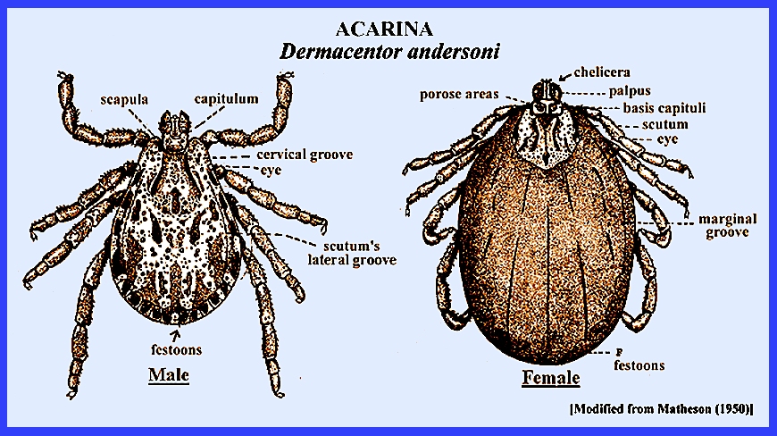
reproduce by parthenogenesis. Being parasites the Ixodidae have developed a simplified
body where the main regions from head to abdomen are contiguous. A scutum or shield is located on the
dorsum whose size and appearance is diagnostic. Females have a smaller shield than males. The eyes occur near the margin of the
anterior portion of the scutum. There
are four pairs of legs in adults, but only three in larvae (See Dermacentor
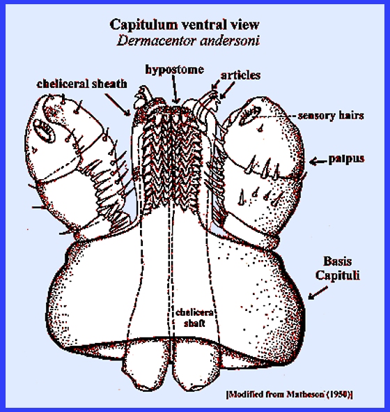
andersoni). A Capitulum near the
anterior portion of the body simulates a true head and is useful in the
identification of species. The basal
portion is the Basis Capituli, which is made u of a broad ring that is
constricted to form the neck, which leads to the anterior opening of the
body. Extending beyond the ring are
the mouthparts that serve for piercing and sucking blood. Palpi arise from the lateroventral margin
of the capituli. The first segment is
typically short followed by second and third longer ones, and a fourth that
is found in a depression on the third segment. The fourth segment regularly has a row of hairs, which are
believed to be sensory. The Hypostome in the shape of an arrow emanates from the
median ventral surface of the basis capituli and extends forward underneath
the mouth opening. Important and
complex cutting organs above the mouth are the Chelicerae, which lacerate
host tissue (See Capitulum). A shield or "scutum" occurs on the top of the
body with variable sizes that aid in species identification (See Dermacentor
andersoni). The
genital opening is situated between the 1st and 2nd or 3rd pair of legs. There are many genera in this family and the majority is
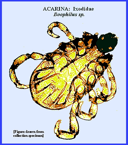
cosmopolitan feeding on mammals, reptiles, amphibians and birds. IMPORTANT IXODID GENERA Genus BOOPHILUS: Boophilus annulatus (Say) is the
cattle tick of North America into Mexico.
It attacks only one host and lays its eggs on the ground. The ticks attach to hosts from the grass
on which they reside. It is important
as a vector of Piroplasma bigemina
or Texas Fever. Genus DERMACENTOR:
Some of the most important
North American ticks are found in this genus, and up-to-date information on
problem species may be sought online.
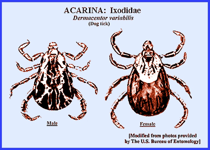
Following are several of the more common species that may be found: Dermacentor variabilis
(Say), the wood- or dog tick, is widely distributed in North America. It has three hosts, with adults preferring
large mammals. The larvae attack
mice. It is an important vector of Rocky Mountain
Spotted Fever virus and Tularemia. Dermacentor andersoni Stiles
bears the name "Rocky Mountain
spotted-fever tick."
It is also distributed widely in Western North America. It is a 3-host tick with a complicated
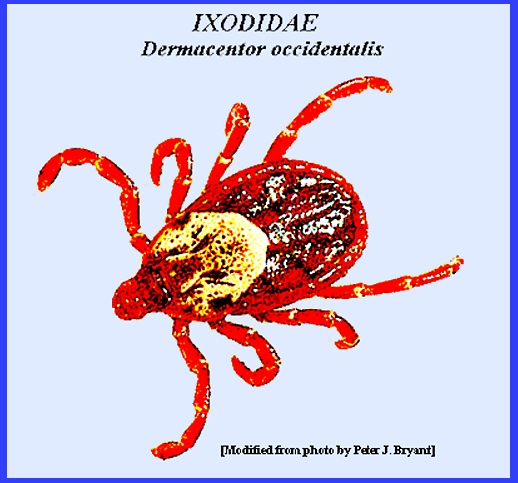
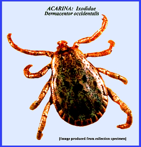
life cycle involving different rodents over two years. Dermacentor occidentalis
Marx ranges along the Pacific coast from California to Oregon where it is
active during the entire year. It is
a 3-host tick that attacks larger domestic mammals as well as humans. A number of rodent hosts are attacked
during its life cycle. On humans the
bite of this tick can be painful and it can transmit tularemia and possibly Rocky Mountain
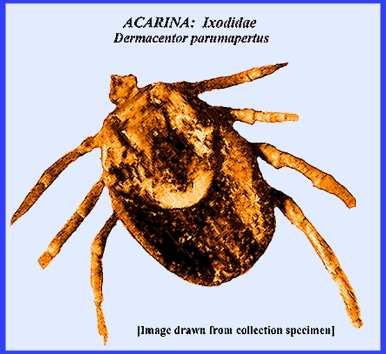
spotted fever. Dermacentor parumapertus
Neum. of Western North America attacks rabbits primarily. Although it does
not frequently encounter humans it is important as a reservoir for the Rocky Mountain
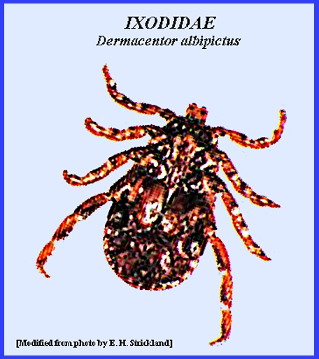
spotted fever virus. Dermacentor albipictus (Pack.)
is known as the Elk Tick. Its
appearance differs from all other ticks in the genus, and it attacks large
game and domestic animals in winter months.
Humans are rarely affected by this species that is distributed all
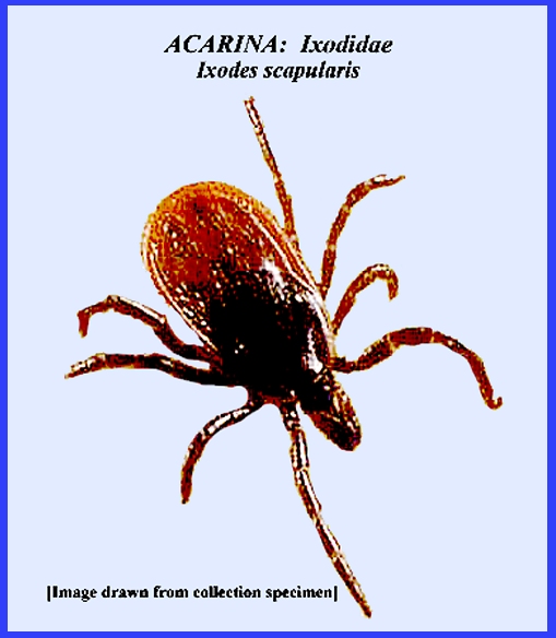
over North America. Genus IXODES: The taxonomy of this genus continues to be revised, but as
of 2017 there were about 55 species known.
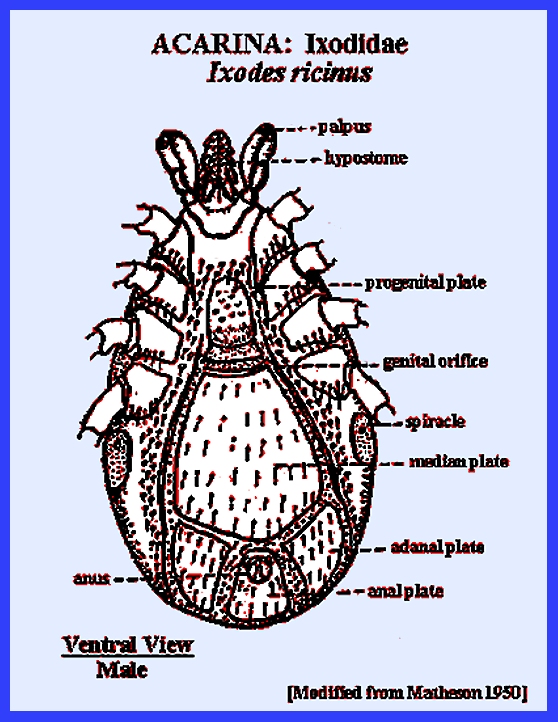
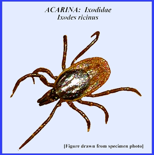
Some important species are noted as follows: Ixodes ricinus (L.) is a
cosmopolitan tick whose hosts include larger mammals and humans. It is a 3-host tick and a vector of the Louping Illness in sheep and
humans. It also vectors Piroplasmosis (Babesia bovis)
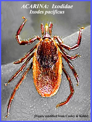
of cattle in Europe. The life cycle covers a whole year. Ixodes pacificus Cooley
& Kohls ranges from Canada to Mexico west of the Cascade Mountains. It is a 3-host tick and may be important
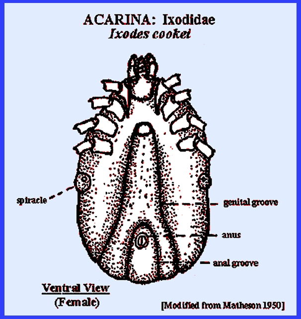
in disease transmission. Ixodes cookei Pack. is common
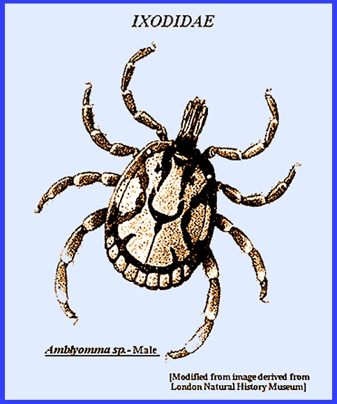
in eastern North America attacking small mammals, cattle and humans. Genus AMBLYOMMA: The genus contains a large number of species that are
common in tropical and subtropical regions of South America and Africa, with
some species found in Eastern North America.
They are very difficult to identify.
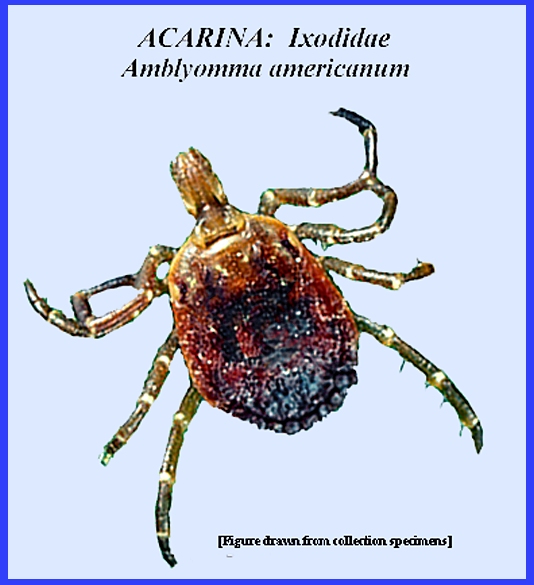
Some important species are noted as follows: Amblyomma
americanum (L.), the Lone Star Tick occurs widely in Eastern
North America. A 3-host tick that
breeds all year long, the larvae and nymphs have a wide host range that
includes birds, mammals and humans.
The bite causes considerable pain followed by persisting
soreness. It has vectored Rocky Mountain
spotted fever in the Central United States, and may also
spread "Q"
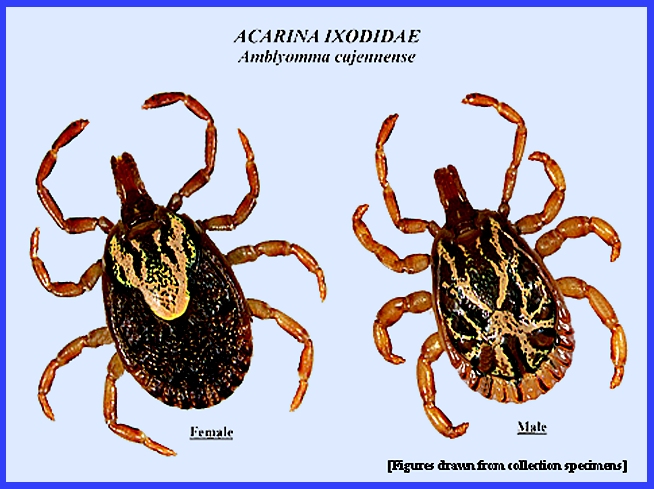
Fever, Bullis Fever and Tularemia. Amblyomma cajennense (Fab.)
ranges from South Texas through Central America and eastern South America. It is a 3-host tick where all stages
attack domestic and wild mammals and humans.
This tick in South America vectors Brazilian
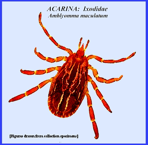
Spotted Fever and Tobia Fever. Amblyomma maculatum
Koch attacks livestock and small wild birds and mammals from Eastern North
America south through South America.
The inflammation resulting from its bite can stimulate attack by
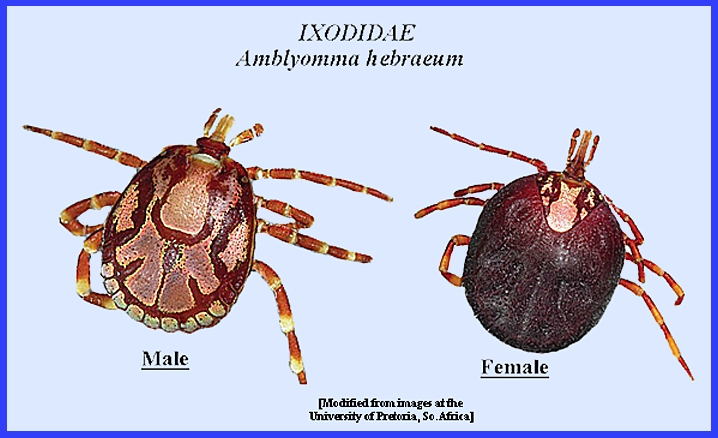
screwworms and the death of domestic animals. Amblyomma hebraeum
Koch, the "Bont Tick" ranges throughout southern Africa. A 3-host tick where all stages attack
humans, domestic and wild animals.
This tick is a vector of "Tick Bite Fever" in
humans and "Heart Water
" of cattle. Genus
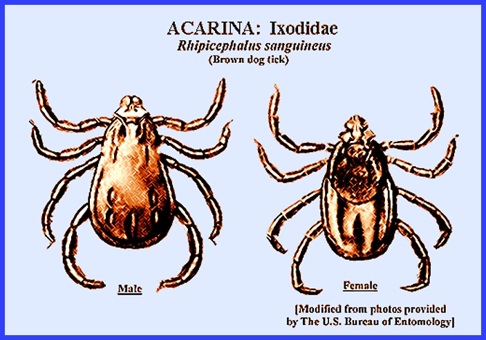
RHIPICEPHLUS: Most species in the Rhipicephalus
genus occur in Africa, with only one cosmopolitan species in North
America.
Rhipicephalus sanguineus
(Latr.), the "Brown Dog Tick" occurs in most of the
tropical and temperate world regions.
It is a 3-host tick with all stages developing on dogs and sometimes
on humans as well. The Rocky Mountain spotted fever virus has
been recovered from this species in North America. It is also a vector of canine piroplasmosis (Babesia canis), and may be involved as
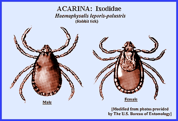
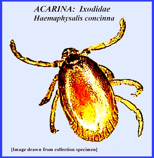
vector of South African Tick-Bite Fever.. Genus HAEMAPHYSALIS: There are only a few species of these small ticks involved
in the transmission of disease. Haemaphysalis
leporis-pulustris (Pack.) is the "Rabbit Tick" that
is an intermediate host for Rocky Mountain spotted fever and
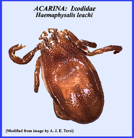
Tularemia. It ranges from Alaska south through South America. Haemaphysalis leachi (Aud.)
has a wide distribution from Africa through Australasia. It vectors "Canine Piroplasmosis" (Babesia
canis) and "Tick Bite
Fever:" in southern Africa.
Haemaphysalis
concinna may be involved as a vector of Russian
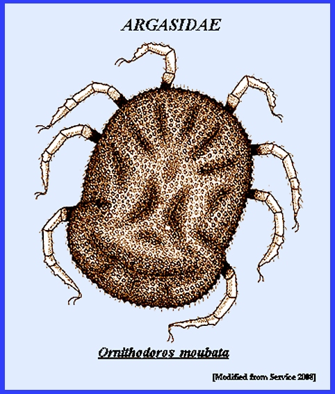
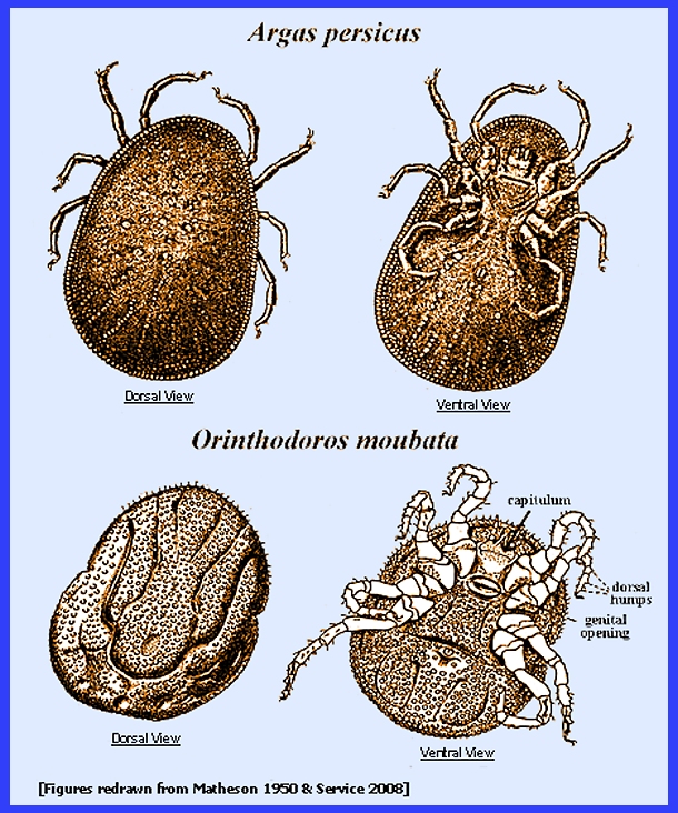
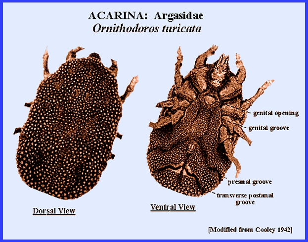
Encephalitis or Tick-Borne Encephalitis. The family differs from Ixodidae
primarily by lacking a dorsal shield or scutum, and the capitulum is on the
ventral surface (See "Examples."). While the underside of the tick resembles the
Ixodidae, the top or dorsal surface is very different, and sclerotization is
minimal. Also, the palpi resemble the
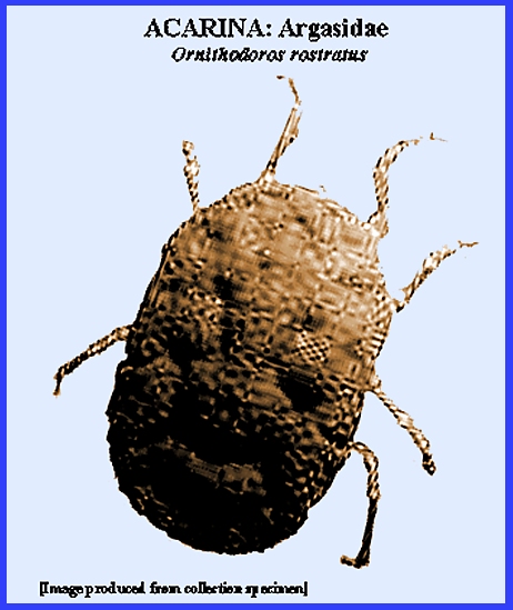
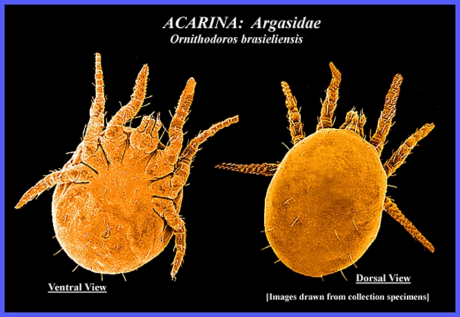
legs of a spider instead of palps. The principal argasid hosts are poultry, domestic animals
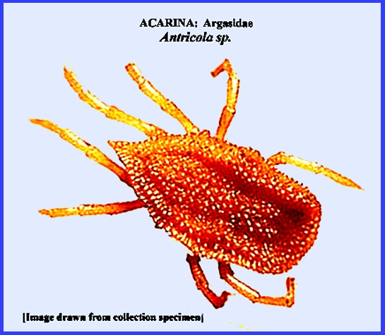
and humans, and they feed primarily at night. Four genera are usually problematic: Argas, Antricola,
Ornithodoros
and Otobius. Genus
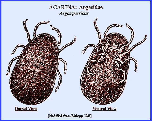
ARGAS: Argas persicus (Oken), the
common fowl tick seeks out domestic fowl, but humans are attacked when in
close contact with the preferred hosts.
This species is cosmopolitan and occurs primarily in structures where
poultry is housed. Argas mianensis
attacks on humans in Iran causes Mianch
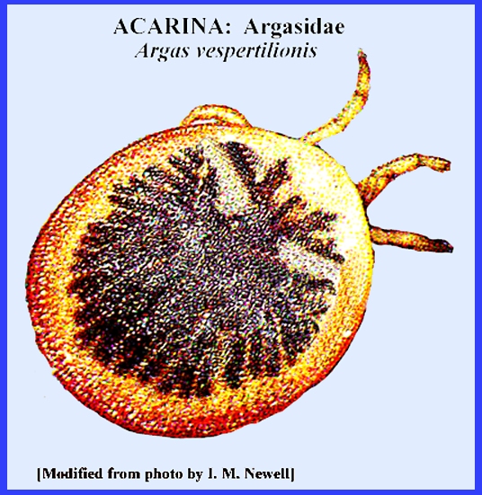
Fever. Argas vespertilionis
(Latr.) attacks bats in northern Europe, and in Africa, India and
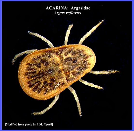
Australia. Argas reflexus (Fabr.) attacks
pigeons in Europe and northern South America, but in North America it is rare
and does not attack pigeons. Argus brumpti
Neum. is a large species (ca. 20 mm.) from East Africa. Genus ANTRICOLA:
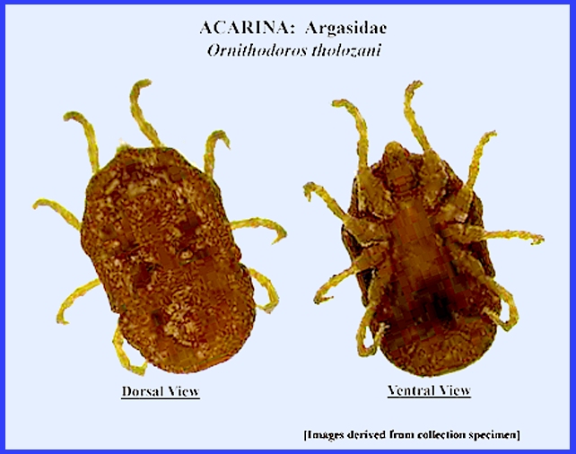
(data being sought). Genus ORNITHODOROS: The genus has a number of species that are important in
transmitting human diseases. Some of
the most important are the following: Ornithodoros moubata
(Murray) prefers humans in all its developmental stages. It also attacks an array of domestic
animals. It occurs in the dry parts
of Africa from Lake Chad to the Red Sea and south to southern Africa and
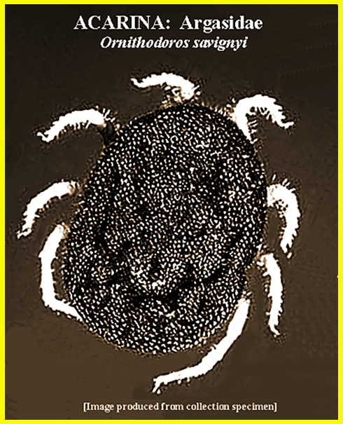
Madagascar. Ornithodoros savignyi
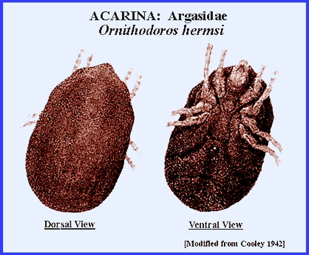
(Aud.) occurs in Africa and east to Arabia and India. It is a vector of relapsing fever. Ornithodoros hermsi Wheeler
is a small tick that occurs at higher elevations in the western United Stages
where it attacks small rodents.
Adults are long-lived and are vectors of relapsing fever. Ornithodoros turicata (Duges)
is a large tick that is abundant in the southwestern and southern United
States and sections of central Mexico.
It attacks an array of domestic and wild animals and humans. It is an important vector of relapsing
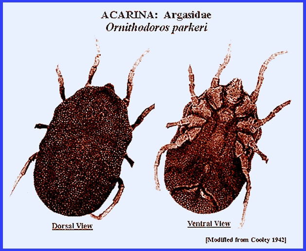
fever. Ornithodoros purkeri
Cooley of the western United States.
It attacks small rodents and humans.
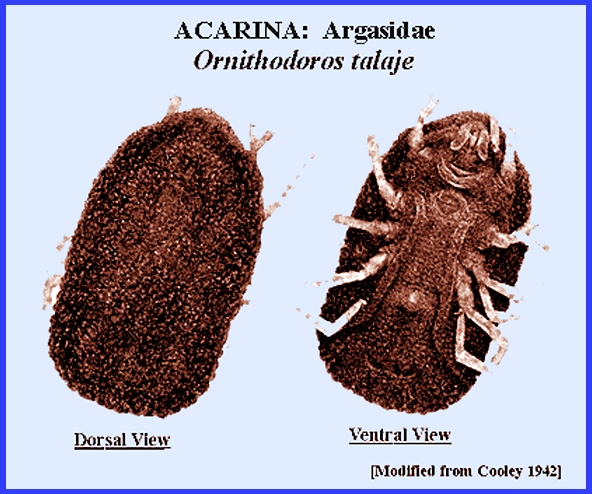
It may be capable of transmitting spotted fevers. Ornithodoros talaje (Guerin-Men.)
is widespread from the southern United States south to Argentina. It attacks an array of mammals, birds and
reptiles and can transmit relapsing fever to humans. Ornithodoros rudus
Karsch) attacks humans primarily in Central and South America, and is a
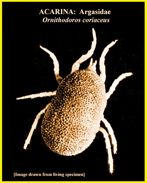
vector of relapsing fever. Ornithodoros coriaceus
Koch is a large tick attacking large mammals and gives a nasty bite to
humans. It is found from California
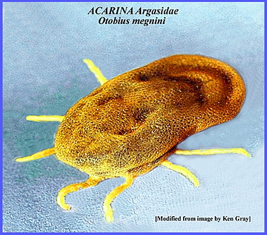
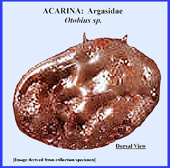
south into Mexico. Genus OTOBIUS: Otobius megnini (Duges)
resides in the ears of horses, cattle and other domestic animals. It is widespread in North America south through
Central and South America. It is also
invaded in southern Africa. Otobius lagophilus whose principal
host is rabbits occurs in the northwestern North America. The bite of ticks can produce serious illness, and the
loss of blood in domestic and wild animals can result in weakness or
death. The bites of some tick species
can result in wounds that are slow to heal and which may become infected or
attract flies that can cause myiasis.
Great care should be taken when removing an embedded tick as crushing
may cause infections. Various
techniques include applying heat to the tick's body or covering it with
adhesive tape. An antiseptic should
be applied to all wounds Tick bites can result in "Tick Paralysis"
especially in young children and domestic animals. A muscular weakness precedes paralysis that can quickly
progress to a loss of leg movement and a spreading to other parts of the body. Removing the tick is essential to avoid
respiratory paralysis and death. As vectors of diseases caused by viruses and pathogens the
role that ticks play has been well known by more than a century. Matheson's (1950) detailed reports of some
of the important diseases are worth noting. Table 1. Tick Species That Inflict Harmful Bites
RELAPSING FEVER.--
A large number of relapsing fevers, caused by Spirochaeta spp. have been
recognized. These fevers are
characterized by repeated attacks that last from 3-5 days. Durations vary from 5-10 days. Causative agents are species of Spirochaeta present in blood,
cerebrospinal fluid and other fluids of the body. During the incubation periods they may not be obvious from the
blood stream although experiments have shown their presence. Ticks are the vectors of various species
of Spirochaeta even though
other arthropods are known to play a role sometimes. The presence of the spirochetes in the
blood stream during the entire infection period is very significant,
especially when prophylactic measures are deployed. Spirochaeta recurrentis (Lebert) was the first
species observed to infest the blood of humans in 1868 by Obermeier, and was
described and named by Lebert in 1874.
Ross (1924) demonstrated that a peculiar fever of West Africa was
caused by a spirochete (S. duttoni),
and that the spirochete was transmitted to humans by the tick Ornithodoros
moubata (Murray). Then
Todd (1914
& 1919) demonstrated that O.
moubata was the vector of the spirochete. The newly hatched offspring of infected
ticks transmitted the disease. Ever
since it has been shown that infection in the tick can pass through the eggs
even to the third generation. Today
many species of spirochetes have been found in the blood of humans and
animals. There are believed to be
more than 15 species or strains occurring in humans alone. Relapsing fevers are now found throughout the world, and
most are passed across generations through the tick eggs. The way transmission of spirochetes occurs
varies for different species. The
ticks obtain the spirochetes while feeding on animal blood that is
infected. In the tick the spirochetes
multiply by transverse fission. They
then invade the tissues and body cavity of the tick. After an infected tick bites a new host,
the spirochetes gain entrance either through the coxal fluid glands, which
eject their secretion of by way of the bite (Davis 1945). ROCKY MOUNTAIN SPOTTED FEVER.--
Ever since settlement certain areas of the North American
west sustained outbreaks of a very fatal disease among the people living
there. The disease was first
recognized around 1890. A high fever
starts the infection followed by arthritic and muscular pains and skin rashes
that begin on the ankles, wrists and forehead, but later may spread over the
entire body. The disease can run a
rapid course, which may end in death after 6-12 days. If the fever drops and the person lives
two weeks recovery is typically quick.
Two strains of the disease exist, a mild and a virulent type. These appear to be present in most of the
geographic regions in which it occurs.
Mortality rates vary from 80 percent for the virulent strain to about
4-6 percent for the mild strain. The
disease bets its name from its area of origin in the Rocky Mountains of North
America. It is not contagious but
highly infectious and transmitted solely by ticks. Warren (1946) suggested that ticks carried the disease and
Ricketts (1906) showed that the disease is mainly an infection of
rodents. Large mammals, except
humans, are not susceptible. The
tick, Dermacentor andersoni,
was found to be the vector for humans.
Wolbach (1916) described and named the parasite Dermacentroxenus rickettsi. For years the disease was known only from
a small area in the Rocky Mountains, but Rumreich et al. (1931) showed it to be present in eastern
North America as well. The dog tick, Dermacentor
variabilis, was found also to vector the disease in the
east. Today we know that the disease
is very widespread throughout North America and down to South America, with Amblyomma
cajennense being the principal vector in the Southern
Hemisphere. There are many different species of ticks now known to
vector the disease over its range. Of
course, transmission can only occur from infected ticks. The incubation in humans after infection
varies from 2-12 days. Vaccines have
been developed for the disease, which can reduce or eliminate symptoms
entirely. TULAREMIA.-- This is a kind of plague in rodents
caused by the bacterium Pasturella
tularensis. It was first
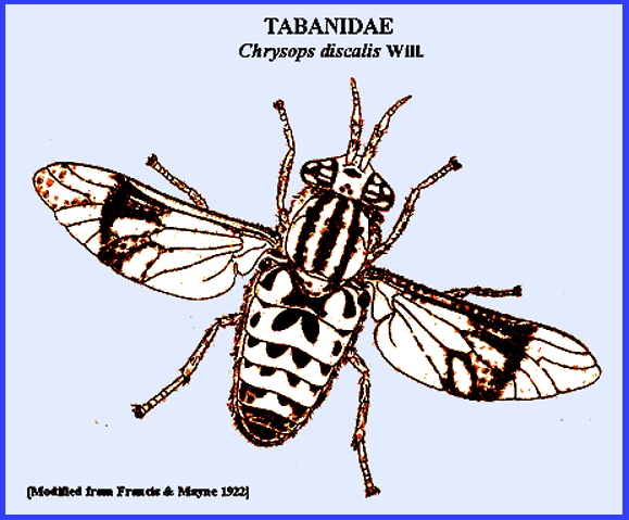
discovered in California rats by McCormack (1921). It was later isolated from squirrels and described by McCormack (1921). Francis (1919, 1920, 1921) showed that "Deer Fly Fever" in humans and the
disease of rodents were identical, being caused by the same organism. It was later named "Tularemia." Tularemia is very infectious to humans being transmitted
by several arthropods through their bites or crushed bodies, or by the feces
and body fluids of rodents. It is
spread throughout North America, Europe, North Africa and Japan. There are many natural reservoid
hosts. Burroughs et al. (1945) listed
44 bird and mammal hosts from different parts of the world. Infection of humans occurs through
contact with reservoir hosts, especially rabbits. The bacterium is very infectious being able to penetrate human
skin. Handling or being around
infected animals can result in infection.
Some arthropods are important as natural reservoirs and also in
transmission to humans. Francis
(1921) first demonstrated that the deer fly, Chrysops discalis can
vector the disease. AUSTRALIAN "Q" FEVER.-- This fever was found to occur among
the meat handlers in Queensland. The
causative organism was found to be Rickettsia
burneti by Burnet & Freeman (1937). A related fever "American
Nine-Mile Fever" was found in Montana in 1938 with the
infectious agent, Rickettsia diaporica,
being isolated from the tick Dermacentor andersoni
(Davis et al. 1939-1943; Cox 1940).
Later it was found that both incitants are identical organisms. The bandicoot rats and other bush animals are reservoirs
in Australia with the tick Haemaphysalis
humerosa probably being
the main vector. Dermacentor
andersoni, Dermacentor occidentalis and
Amblyomma americanum
are vectors in North America.
Infection of this disease is also very common by inhalation around
infected animals and meat. Q Fever was a serious problem among troops during World
War II in Europe with different strains being involved. COLORADO TICK FEVER.-- Occurring in the Rocky Mountains of
North America, the disease is associated with the bite of Dermacentor andersoni. A rash does not occur and the fever is a
remittent type with rare fatality. BULLIS FEVER.--
The fever was reported this disease from Texas in 1943 and
was isolated from troops during World War II. The vector was reported as Amblyomma americanum
because of its frequency around infected individuals (Matheson 1950). TICK TYPHUS.--
This disease has been found from different parts of the
world. The tick Dermacentor nuttalli Olenev was reported
as a vector in Russia, with rodents being reservoirs. It seems to be spread in India, East
Africa and the Americas, but detection is not always certain. BOUTONNEUSE FEVER.--
First reported from Tunisia by Conor & Bruch (Matheson
1950) the disease is known to be widespread in Europe and Ethiopia. The causative agent is Rickettsia conori with the tick vector Rhipicephalus
sanguineus. Rodents
and domestic pets serve as reservoirs for the disease. SOUTH AFRICAN TICK BITE FEVER.-- Closely related to spotted fevers
caused by rickettsia, this disease was called Rickettsia rickettsi conori or a strain of R. r. pijperi in southern Africa. The reservoirs are dogs and the vectors
the dog ticks, Haemaphysalis leachi,
Amblyomma hebraeum
and possibly Rhipicephalus
sanguineus. However,
only the larval stage was believed to transmit the pathogen. It is usually associated with tick bites
followed by a sore and lymphadenitis. Transovarial transmission is also possible
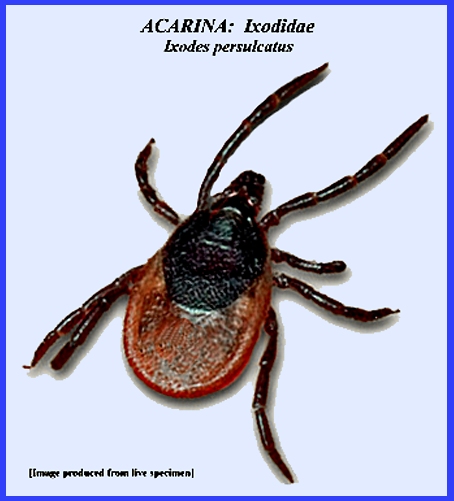
by the tick. RUSSIAN TICK BORNE ENCEPHALITIS.-- This is one of the very few
encephalitis of humans that is transmitted by ticks. The distribution is primarily Russia all
the way to the Far East where there is virgin forest. The tick Ixodes persulcatus
Schulze is a principal vector, although Dermacentor
silvarum, Haemaphysalis concinna and
H. japonica are
suspected.. Transmission to humans
that spend time in forests of the distribution area. Infection occurs in spring and summer,
with the first from the overwintering ticks and the second from the young
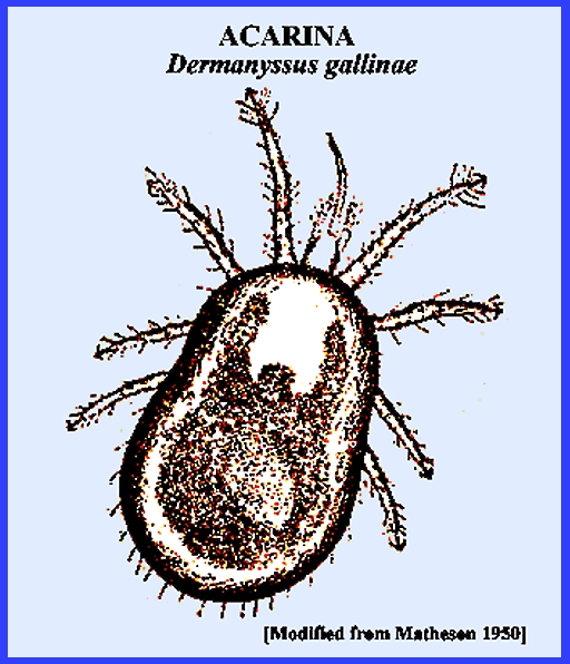
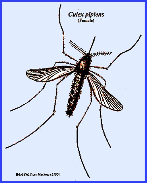
that hatch from eggs deposited in springtime. SAINT LOUIS ENCEPHALITIS.-- This virus is transmitted by the
Chicken Mite, Dermanyssus gallinae,
and chickens serve as reservoirs for the disease. The mosquito, Culex pipiens, and a
number of other mosquito species that attack humans acquire the virus from
chickens and thereby become infectious for other animals including humans. ANIMAL DISEASES TRANSMITTED.-- Many diseases of domestic and wild
animals are transmitted by ticks, including
Anaplasmosis, East Coast Fever, Fowl Spirochetosis, Heartwater of Sheep & Cattle. Louping Ill Disease. Nairobi Sheep Disease. Piroplasmosis, Red Water Fever, Texas Fever, etc. As these hosts may serve as disease
reservoirs, close contact with the infected animals may pose a hazard for
humans as well (Please check the Internet for more information on animal
diseases). Avoiding tick habitats and the diseases they carry is
about the best way to avoid bites and infection. Traditionally various dips and sprays have been used for
domestic animals. There are also
vaccinations available for some diseases, and consulting a physician is
advised for the latest treatments.
Key References: <medvet.ref.htm> Bishopp. R. C. 1935. Ticks and the role they play in the transmission of
disease. Rept. Smithsonian Inst. , p.
389-406. Bowman, A. S. & P. A.
Nuttall (eds.). 2004. Ticks: biology, disease &
control. Parasitology 129: S1-S450 Brumpt, E. 1927.
Précis de paraaitologie. 4th ed. Paris, France. Burnet, F. F. & M.
Freeman. 1937. Experimental studies on the firus of
"Q" fever. Med. Jl. Australia
2: 299-305. Burroughs, A. R.
et.al. 1945. A field Study of latent tularemia in
rodents with a list of all known naturally infected vertebrates. Jl. Inf. Dis. 76(2): 115-19. Camicas, J. L., J. P. Hervy, F. Adam & P. C.
Morel. 1998. The Ticks of the World (Acarida, Ixodida):
Nomenclature, Described Stages, Hosts, Distribution. Editions de l'ORSTOM, Paris. Cox, H. R. 1940.
Rickettsia diaporica and
American "Q" fever. Amer.
J. Trop. Med. 20: 463-69. Cunha, B. A. (ed.). 2001.
Tickborne Infectious Diseases:
Diagnosis & Management. Marcel Dekker, NY. Davis, Gordon E.
1939. Ornithodoros parkeri; distribution and
host data; spontaneous infection with relapsing fever spirochetes. U.S. Pub. Hlth. Repts. 54: 1345-1349. Davis, Gordon E.
1940. Bacterium tularense; its persistence in
the tissues of the argasid ticks Ornithodoros
turicata and O. parkeri. U. S. Pub. Hlth Repts. 55: 676-80. Davis, Gordon E. 1941.
Ornithodoros parkeri
Cooley: observations on the biology
of this tick. J. Parasit. 27: 425-33. Davis, Gordon E. 1942.
Tick vectors & life cycles of ticks. IN: ymposium on relapsing fever in the Americas. Amer. Assoc. Adv. Sci., Pub. 18: 67-76. Davis, Gordon E.
1943. American Q fever;
experimental transmission by the argasid ticks Ornithodoros moubata and O.
hermsi. U.S. Publ. Hlth. Repts. 58: 984-87. Davis, Gordon E.
1943. Experimental
transmission of the rickettsiae of spotted fevers of Brazil, Colombia and the
United States by the argasid tick, Ornithodoros
parkeri. U.S. Publ. Hlth
Repts. 58: 1201-1208 Davis, Gordon E. 1943.
Studies of the biology of the argasid tick, Ornithodoros nicollei Mooser. J. Parasit. 29: 393-95. Davis, Gordon E.
1943. The tick Ornithodoros rudis as a host to the
rickettsiae of the spotted fevers of Colombia, Brazil and the United States. U.S. Publ. Hlth. Repts. 58: 1016-1020. Dumler, J. S. & D.
H. Walker. 2005. Rocky mountain spotted fever: changing
ecology and persisting virulence. New
England J. Med. 353: 551-53. Francis, Edward et
al. 1922. Tularaemia: a new disease of man. U. S. Pub. Hlth. Sev. Hyg. Lab. Bull. 130. Francis, Edward.
1927. Microscopic changes of
tulraemia in the tick, Dermacentor
andersoni, and the bedbug, Cimex
lectularius. U.S. Publ. Hlth. Repts. 42: 2763-2772. Francis, Edward. 1929.
Arthropods in the transmission of tularaemia. Trans. 4th Internat. Cong. Ent. 2: 929-944. Francis, Edward.
1942. The longevity of fasting
and non-fasting Ornithodoros turicata
and the survival of Spirochaeta
obermeieri within them. In:
Symposium on relapsing fever in the
Americas. Amer. Assoc. Adv. Sci. Pub.
18: 85-88 Gammons, M. & G.
Salam. 2002. Tick removal. Amer. Fam. Physician 66:
643-45. Gothe, R., K. Kunze
& H. Hoogstraal. 1979. The mechanisms of pathogenicity in the
tick paralyses. J. Med. Ent. 16: 357-69. Gray, J. S., O. Kahl, R.
S. Lane & G. Stanek. 2002. Lyme Borreliosis: Biology, Epidemiology
& Control. CABI, Wallingford,
England Hoogstraal, H. 1966.
Ticks in relation to human diseases caused by viruses. Ann. Rev. Ent. 11: 261-308. Hoogstraal, H. 1981.
Changing patterns of tickborne diseases in modern society. Ann. Rev. Ent. 26: 75-99. Kisinza, W. N., P. J.
McCall, H. Mitani, A. Talbert & M. Fukunga. 2003. A newly
identified tick-borne Borrellia
species and relapsing fever in Tanzania.
Lancet 362: 1283-84. Klompen, J. S. H., W. C.
Black, J. E. Keirans & J. H. Oliver.
1996. Evolution of ticks. Ann. Rev. Ent. 41: 141-61. Lane, R. S., I. Piesman & W. Burgdorfer. 1991.
Lyme borreliosis: relation of its causative agent to its vectors and
hosts in North America and Europe. Ann. Rev. Ent. 36: 587-609. Lawrie, C. H., N. Y. Uzcategui, E. A. Gould & P.
A. Nuttall. 2004. Ixodid and argasid tick species & West
Nile virus. Emerging Infectious Diseases 10: 653-57. Mail, G. A. & J.
D. Gregson. 1938. Tick paralysis in British Columbia. J. Canad. Med. Assoc. 39: 532-537. Matheson, R. 1950. Medical Entomology. Comstock Publ. Co, Inc. 610 p. McCormack, P. D. 1921.
Paralysis in children due to the bites of wood ticks. J. Amer. Med. Assoc. 77: 260-63. Needham, G. R. & P.
D. Teel. 1991. Off-host physiological ecology of ixodid
ticks. Ann. Ref. Ent.
36: 313-352. Nuttall, G. H. F.
1908. The Ixodoidea or ticks,
spirochaetosis in man and animals, piroplasmosis. The Harben Lectures.
Roy. Inst. Pub. Hlth. Nuttall, G. H. F. &
C. Warburton. 1908. A monograph of the Ixodoidea, Part 1. Argasidae. Parola, P. & D. Raoult. 2001.
Tick-borne typhuses. IN: The Encyclopedia of Arthropod-transmitted
Infections of Man & Domesticated Animals. CABI: 516-524. Patton, W. S. & F.
W. Cragg. 1913. A textbook of medical entomology. Calcutta & London. Patton, W. S. & A. M. Evans. 1929-1931. Insects, ticks, mies and venomous animals of medical and
veterinary importance. Part I. Medical; Part 2, Public Health. Croydon, England. Ricketts, H. T.
1906. The transmission of
Rocky Mountain spotted fever by the bite of the wood tick (Dermacentor occidentalis). J. Amer. Med. Assoc., 47: 358 Ross, I. C.
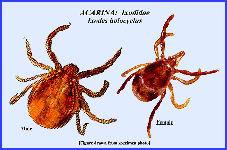
1924. The bionomics of Ixodes
holocyclus Neumann, with a redescription of the adult & nymphal
stags & a description of the larvae Rumreich, A., R. E.
Dyer & L. F. Badger. 1931. The typhus-Rocky Mountain spotted fever
group. U.S. Pub. Hlth. Repts.
49: 470-80 Sauer, J. R. & J. A.
Hair. 1986. Morphology, Physiology & Behavioral Ecology of Ticks. Ellis Horwood & Wiley, New York. Schuster, R. & P. W.
Murphy. (eds.). 1991. The Acari: Reproduction, Development &
Life History Strategies. Chapman
& Hall, London. Service, M. 2008.
Medical Entomology For Students.
Cambridge Univ. Press. 289 p Sonenshine, D. E. 1993.
Biology of Ticks, Vol. 2.
Oxford Univ. Press. Sonenshine, D.
E. 2006. Tick pheromones and their use in tick control. Ann. Rev. Ent. 51: 557-580. Sonenshine, D. E., R. S. Lane & W.
L. Nicholson. 2002. Ticks (Ixodida). IN: Medical & Vet.
Ent.. Academic Press, Amsterdam. pp. 517-558. Steere, A, J. Coburn
& L. Glickstein. 2005. Lyme borreliosis. IN:
Tick-Borne Diseases of Humans.
ASM Press, Washington D.C. Todd, J. L. 1914.
Tick paralysis. J. Parasit.
1: 55-64. Todd, J. L. 1919.
Tick caused paralysis. J.
Canad. Med. Assoc. 9: 994-96. Warren, Joel. 1946.
Epidemic encephalitis in the Far Est.
Amer. J. Trop. Med. 26:
417-436. Wolbach, S. B. 1916.
The etiology of Rocky Mountain spotted fever. J1.. Med. Res.,34:
121-126. |