|
1. The larva
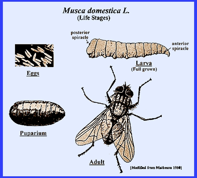
like that of the housefly (Fig. 1);
body slender, cylindrical &
tapers anteriorly & more truncate posteriorly _ _
2
Larva
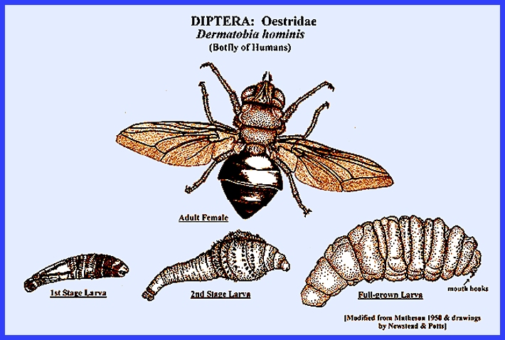
is large, stout and resembles that of a warble, Hypoderma sp.; larva cylindrical or
flattened, depressed or pear-shaped
(Fig. 6)_ _
_ _ _ _ _ _ _ _ _ _ _ _ _ _ _ _ _ _
_ _ _ _ _ _ _ _ _ _ _ _ _ _ _ _ _ _ _ _ _ _ _ _ _ _ _ _ _ _ _ _ _
_ _ _ _ _ _ _ _ 9
Larva
has spiny or fleshy lateral, dorsal or terminal processes (Fig. 2) _ _ _ _ _ _ _ _ _ _ _ _ _ _ _ _ _ _ _ _
_ _ _ _ _ _ _ _ _ 16
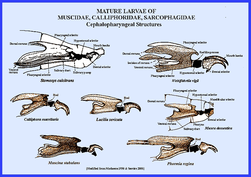
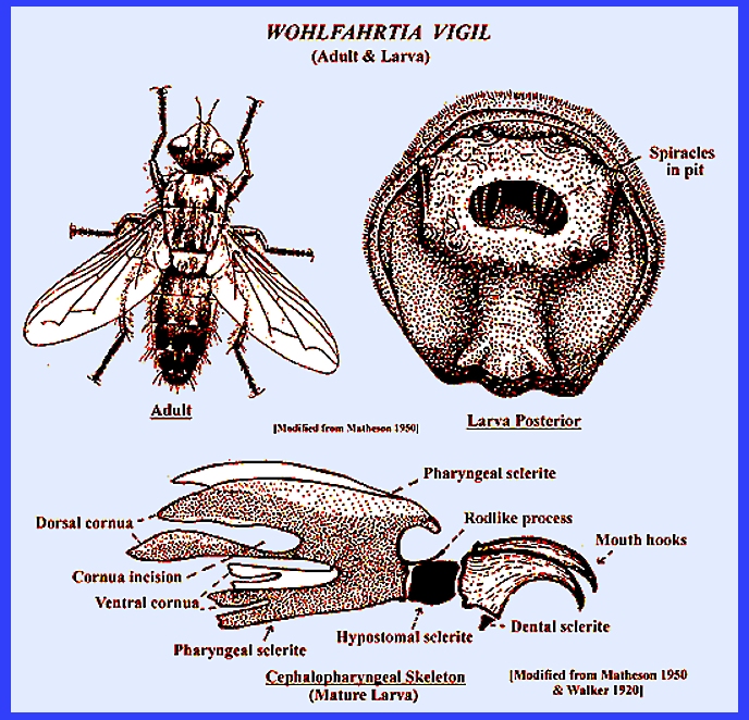
2. The last obvious segment (anal) has a
deep concavity holding the spiracles (Fig. 3);
each dorsal cornua of the pharyngeal
sclerite has a deep
posterior incision (Fig. 3) _ _ _ _ _ _ _ _ _ _ _ Sarcophagidae (Wohlfahrtia spp., Sarcophaga spp.)
The
last segment does not have a deep concavity; spiracles are almost flush with
the posterior face of anal segment; dorsal
cornua of the pharyngeal sclerite does not have an incision _ _ _ _ _ _ _ _ _ _ _ _ _ _ _ _ _ _ _ _
_ _ _ _ _ _ _ _ _ _ _ _ _ _ _ 3
3. Openings of posterior spiracles
sinuous, short or a bit long; button area
usually deep in the peritreme (Fig. 4 -#8
& #9)_
_ _ _ _ _ _ _ _ _ _ _
_ _ _ _ _ _ _ _ _ _ _ _ _ _ _ _ _ _ _ _ _ _ _ _ _ _ _ _ _ _ _ _ _ _ _ _ _ _
_Muscidae (part) _ _ _ _ _ _ _ 6
Openings of posterior spiracles long,
slender, quite parallel & directed downward (Fig. 4 #5)
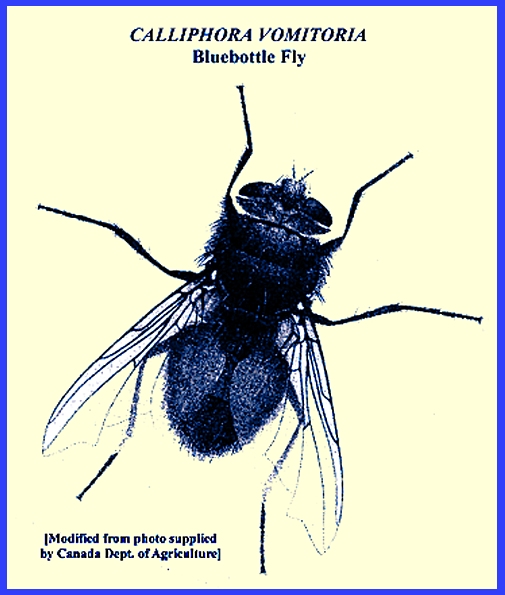
_ Calliphoridae (part) 6
4. Posterior spiracles are D-shaped; there
are 3 sinuous slits in each spiracle's plate (Fig. 4 #8) _ _ _ _ _ Musca domestica
Posterior spiracles are not D-dhaped;
they are rounded or a bit irregular _ _ _ _ _ _ _ _ _ _ _ _ _ _ _ _ _ _ _ _
_ _ _ _ _ _ _ _ 5
5. Spiracle openings slightly curved &
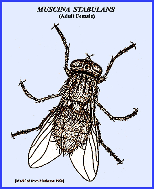
surrounded by large, dense peritreme (Fig. 4 #4) Muscina stabulans; Muscina spp.
Spiracle openings are S-shaped with a dense peritrreme; a button is
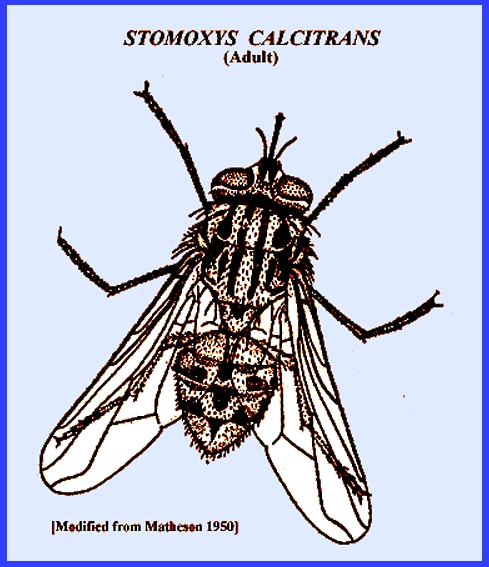
located in the center (Fig. 4 #9) _ Stomoxys
calcitrans
6. The peritreme of posterior spiracles is complete
and with a distinct button (Fig. 4 #5
& #7)_
_ _ _ _ _ _ _ _ _ _ _ _ _ _ _ _ _
_ _ _ _ _ _ _ _ _ _ _ _ _ _ _ _ _ _ _ _
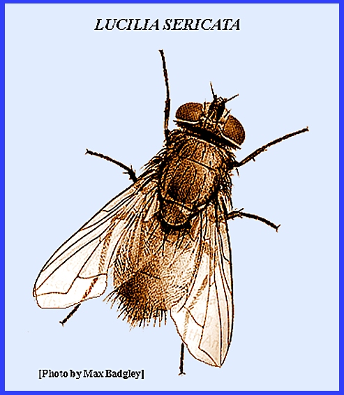
_ _ _ _ _ _ _ _ _ _ _ _ _ _ _ _ _ Calliphora spp., Lucilia spp., Cynomopsis
spp.
The peritreme is not
complete and the button not pronounced and barely visible or in a thinner
area of the peritreme _ _ _ 7
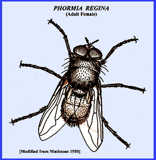
7. Posterior spiracles with button located
in a thinner area of the peritreme (Fig. 195 #6) _ _ _ _ _ _ _ _ _ Phormia regina
Posterior spiracles without a button are not indicated _ _ _ _ _ _ _
_ _ _ _ _ _ _ _ _ _ _ _ _ _ _ _ _ _ _ _ _ _ _ _ _ _ _ _ _ _ _ _ 8
8. The trunks of the trachea that extend
from the posterior spiracles are not pigmented (Fig. 4 #2)
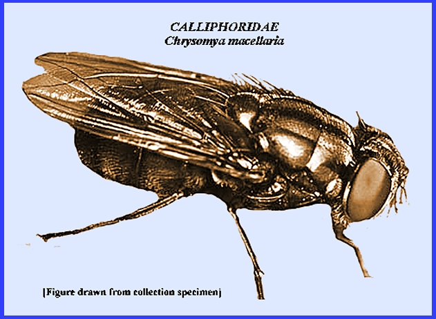
_ Callitroga macellaria
( Also in genera Chrysomya
or Cochliomyia)
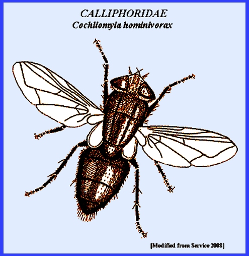
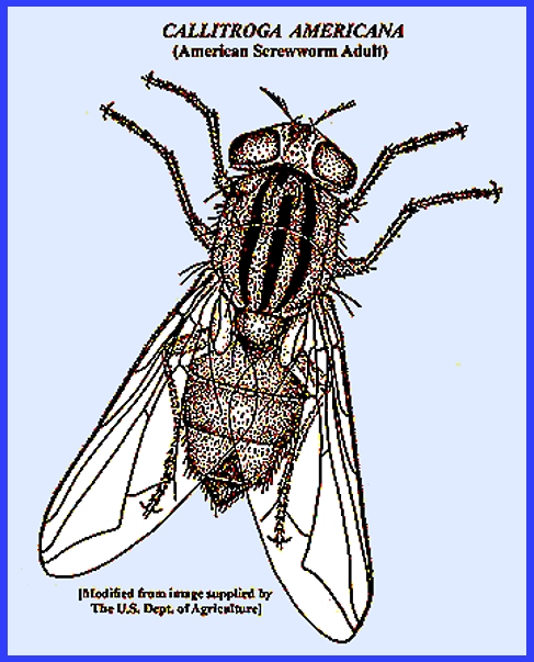
The
trunks intensely pigmented for most of their length (Fig. 187) (The true American screwworm) _ Callitroga
americana
( Also in genera Chrysomya
or Cochliomyia)
9. Each posterior spiracle has 3 distinct
slits (Fig. 4 #1,
#2,
#3) _ _ _ _ _ _ _ _ _ _ _ _ _ _ _ _ _ _
_ _ _ _ _ _ _ _ _ _ _ _ _ 10
Each
posterior spiracle has many small openings that are without pronounced
slits (Fig. 5) _ _ _ _ _ _ _ _ _ _ _ _ _ _ _ _ 13
10. The larvae are pear-shaped (Fig. 6) and
greatly spined; spiracular openings
are straight and located in a deep crater _ _ _ _ _
_ _ _ _ _ _ _ _ _ _ _ _ _ _ _ _ _ _ _ _ _ _ _ _ _ _ _ _ _ _ _ _ _ _
_ _ _ _ _ _ _ _ _ _ _ _ _ _ _ _ _ _ _
Dermatobia hominis
Larvae
are egg-shaped; spiracular openings are bent at the middle and located in a
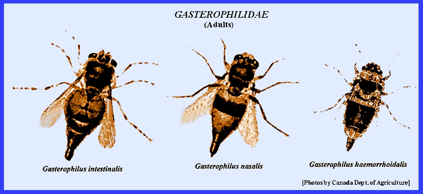
shallow crater _ _ _ _ Gasterophilidae
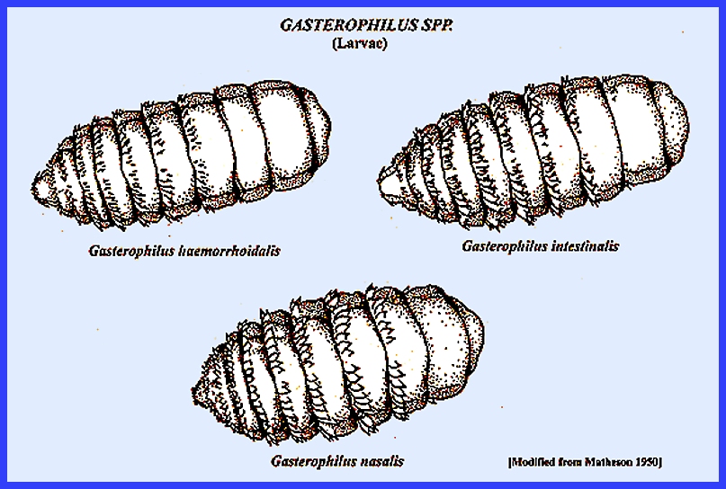
11. Spines located on segments' anterior margins
stout & arranged in single row (Fig. 7) _ _ _ Gasterophilus nasalis 11
The spines
are arranged in a double row _ _ _ _ _ _ _ _ _ _ _ _ _ _ _ _ _ _ _ _ _ _ _
_ _ _ _ _ _ _ _ _ _ _ _ _ _ _ _ _ _ _ _ _ _ 12
12. Spines on the anterior margins are
small, tapering to a fine point; there are no spines on the dorsum of
segment #11 and the
middle of segment #10 (Fig. 7) _ _
_ _ _ _ _ _ _ _ _ _ _ _ _ _ _ _ _ _ _ _ _ _ _ _ _ _ Gasterophilus haemorrhoidalis
The
anterior row of spines are thick, more blunt and larger; spines exist on
the dorsum of segment #1 and sevral on each side
of the dorsum of segment 11
(Fig. 7) _ _
_ _ _ _ _ _ _ _ _ _ _ _ _ _ _ _ _ _ _ _ _ _ _ _ _ _ _ _ _ _ _ _ _ _ Gasterophilus intestinalis
13. Mouth hooks are not well developed _ _ _ _ _ _ _ _ _ _ _ _ _ _ _ _ _ _ _ _
_ _ _ _ _ _ _ _ _ _ _ _ _ Hypodermatidae 14
Mouth hooks are well developed
_ _ _ _ _ _ _ _ _ _ _ _ _ _
_ _ _ _ _ _ _ _ _ _ _ _ _ _ _ _ _ _ _ _ _ _ _ _ _ _ _ _ _ _ _ _ _ 15
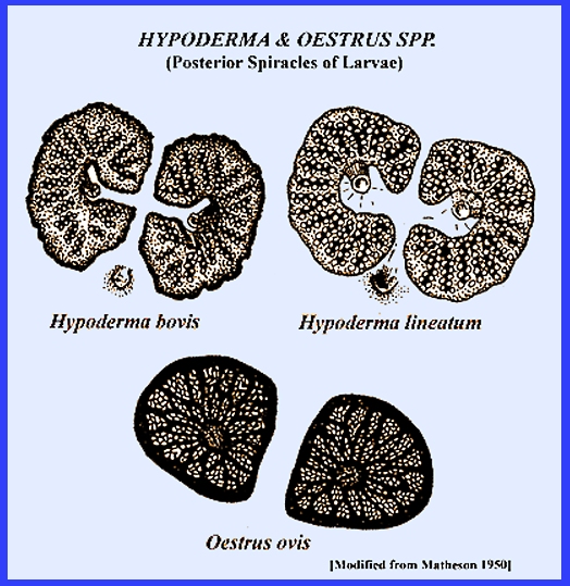
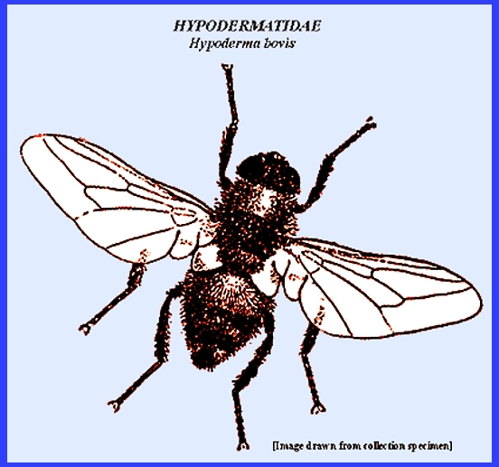
14. The posterior spiracles have their
stigmatal plate profoundly cracked and pointing like a funnel toward the
button (Fig. 8)_
_ _ _ _ _ _ _ _ _ _ _ _ _ _ _ _ _ _ _ _ _ _ _ _ _ _ _ _ _ _ _ _ _ _
_ _ _ _ _ _ _ _ _ _ _ _ _ _ _ _ _ _ _ _ Hypoderma bovis
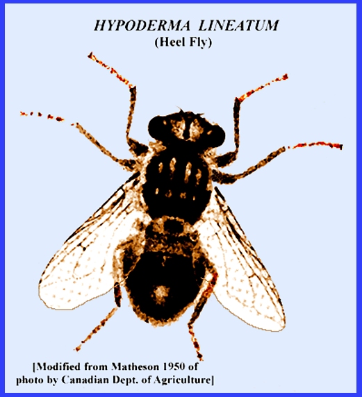
The
posterior spiracle has a stigmatal plate that is less cracked pointing
toward the button (Fig. 8) Hypoderma lineatum
15. Mouth hooks shaped like a horn; the
body has poorly developed spines; the posterior spiracles are heavily
sclerotized with
a button in the center and forming part of the plate (Fig. 8) _ _ _ _ _ _ _ _ _ _ _ _ _ _ _ _ _ _ _ _
_ _ _ _ _ Oestrus ovis
Mouth
hooks are not so hard; the body is thickly set with spines or hard scales (Fig. 9); posterior
spiracles are divided into
plates _ _ _ _ _ _ _ _ _ _ _ _ _ _ _ _ _ _ _ _ _ _ _ _ _ _ _ _ _ _ _
_ _ _ _ _ _ _ _ _ _ _ _ _ _ _ _ _ _ _ _ _ _Cynomopsis spp.
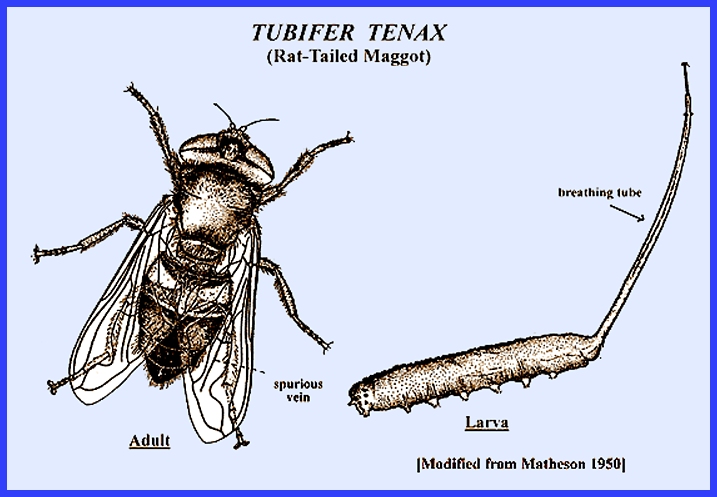
16. Larvae are cylindrical, stout and with a
long posterior tubular extendable process (Fig. 10) _
_ _ _ _ _ _ _ _ Tubifera spp.
Larvae
are not cylindrical and a posterior extended process is absent; lateral and
dorsal fleshy processes or spines are
present _ _ _ _ _ _ _ _ _ _
_ _ _ _ _ _ _ _ _ _ _ _ _ _ _ _ _ _ _ _ _ _ _ _ _ _ _ _ _ _ _ _ _ _ _ _ _ _
_ _ _ _ _ _ _ _ _ _ _ _ _ 17
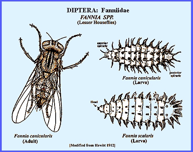
17. The fleshy posterior processes are
somewhat feathery (Fig. 11) _ _ _ _ _ _ _ _ _ _ _ _ _ _ _ _ _ _ _ _
_ Fannia scalaris
The
fleshy posterior processes are simple and shaped more likes spines (Fig. 11) _ _ _ _ _ _ _ _ _ _ Fannia
canicularis
|