File: <tabanidaekey.htm> <Medical
Index> <General Index> Site Description Glossary <Navigate to Home>
|
Insecta: Diptera TABANIDAE KEY (Horseflies &
Deerflies, etc.) (Contact)
Please CLICK on underlined links to view images or to navigate
within the key: To Search for Subject Matter use Ctrl/F [See: Tabanidae Details] Among the Tabanidae there
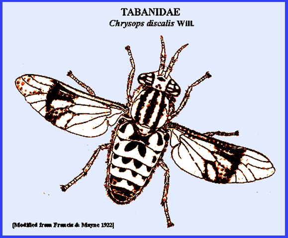
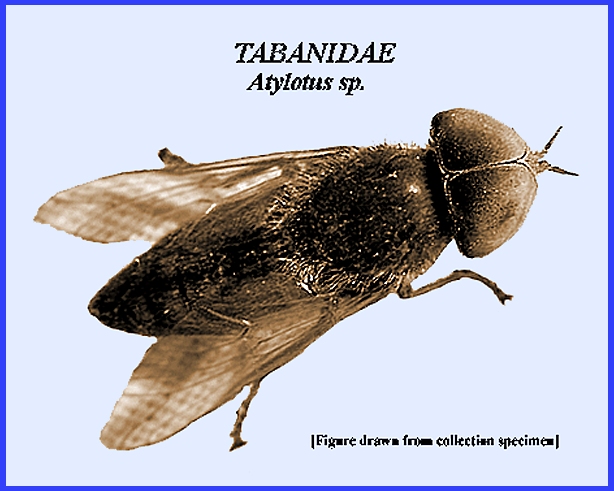
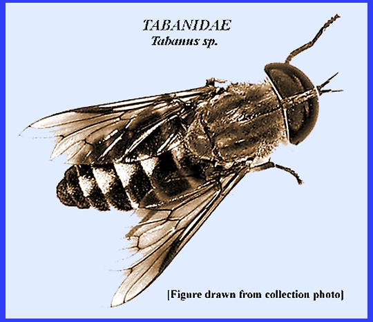
is a considerable size range. Many of
the larger species are built heavily (Fig. 1) and are known for their
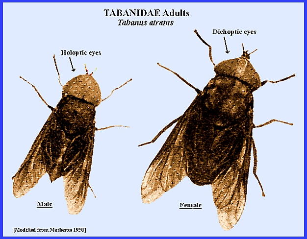
biting habits (Fig. 2). Their color varies from brown to black
with some species having green stripes on their abdomen. The head is large and shaped like one-half
of a circle. The sexes are
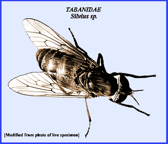
distinguished by males having holoptic eyes and females dichoptic eyes (Fig. 1). Antennae are not very long but somewhat wide
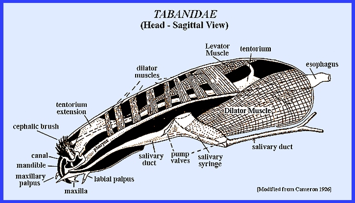
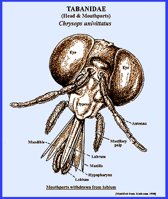
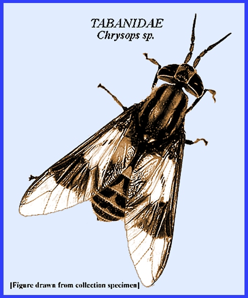
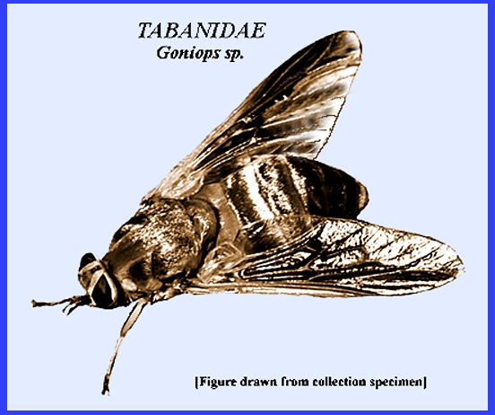
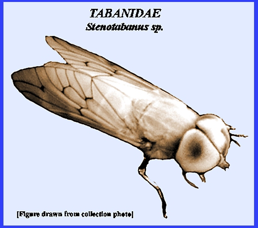
(Fig. 3 & Fig. 4). This is a large family
with over 62 genera. Two subfamilies,
Pangoniinae and Tabaninae
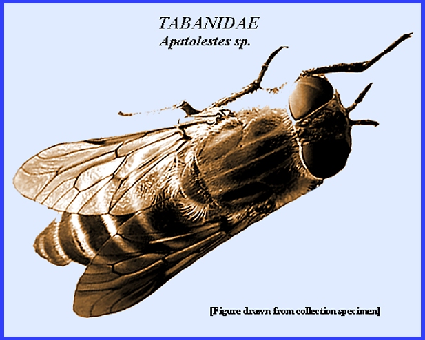
include eleven medically important genera, which are Apatoletes, Atylotus,
Chrysops,
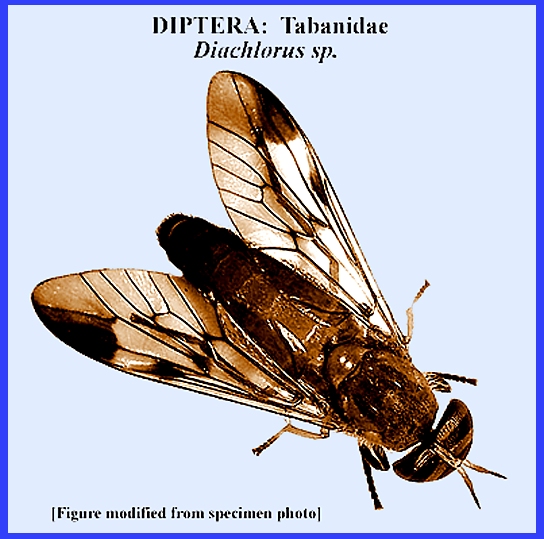
Diachlorus,
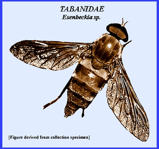
Esenbeckia,
Goniops,
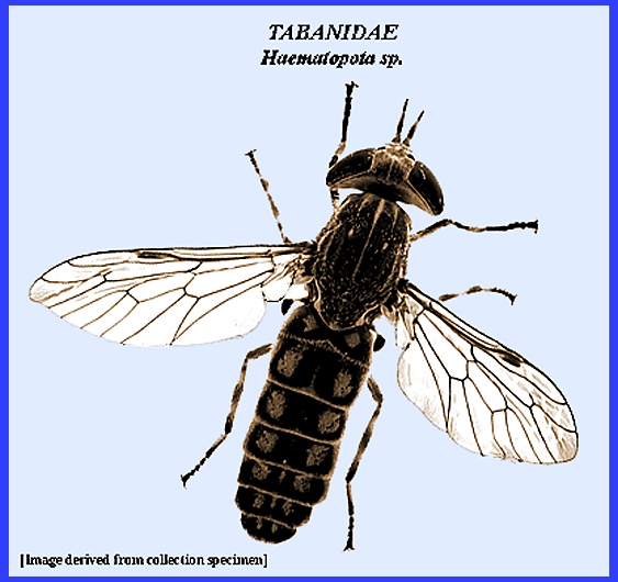
Haematopota,
Silvius,
Stenotabanus,
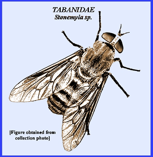
Stonemyia
and Tabanus. The present key separates these 11 genera genera (unmarked images created from collection specimens).
_ _ _ _ _ _ _ _ _ _ _ _ _ _
_ _ _ _ _ _ Key References: <medvet.ref.htm> <Hexapoda> Anderson, J. F. 1985.
The control of horse flies and deer flies (Diptera: Tabanidae). Myia 3:
547-98. Anthony, D. W. 1962.
Tabanids as disease vectors. IN: Biological Transmission of Disease
Agents. Academic Press, NY. p.
93-107. Cheke, R. A., J. Mas
& J. F. Chainey. 2003. Potential vectors of Ioiasis and other
tabanids on the island of Bioko, Equatorial Guinea. Med. Vet. Ent. 17: 221-3. Chippaux, J. P., B.
Bouchite, M. Demanov, I. Morlais & G. LeGoff. 2000. Density and
dispersal of the Iolasis vector Chrysops dimidiata in southern Cameroon. Med. & Vet. Ent.
14: 339-44. Foil, L. D. 1989.
Tabanids as vectors of disease agents. Parasitol. Today 5:
88-95. Francis, E. & B.
Mayne. 1922. Experimental transmission of tularemia by
flies of the species Chrysops discalis. U.S. Pub. Hlth. Svc. Bull. 130: 8-16. Matheson, R. 1950. Medical Entomology. Comstock Publ. Co, Inc. 610 p. Noireau, F., A. Nzoulani, D. Sinda &
A. Itoua. 1990. Transmission indices of Loa loa in the Chaillu Mountains,
Congo. Amer. J. Trop. Med. 43:
382-8. Anthony, D. W. 1962.
Tabanids as disease vectors.
IN: Biological Transmission of Disease Agents. Acad. Press. pp 93-107. Service, M. 2008.
Medical Entomology For Students.
Cambridge Univ. Press. 289 p Legner, E. F. 1995. Biological control of Diptera of medical and veterinary
importance. J. Vector Ecology 20(1):
59-120. Legner, E. F. 2000.
Biological control of aquatic Diptera. p. 847-870.
Contributions to a Manual of Palaearctic Diptera, Vol. 1, Science Herald, Budapest. 978 p. Thomson, M. C., V.
Obsomer & J. Kamgno et al.
2004. Mapping the distribution
of Loa loa in Cameroon in support of the African Programme for Onchocerciasis Control. Filaria J. 3: 7. |