File: <sarcoptoidea.htm> <Medical
Index> <General Index> Site Description <Navigate to Home>
|
Acarina SARCOPTOIDEA (Parasites of
Birds, Mammals & Insects) (Contact) Please CLICK on
Image & underlined links to view:
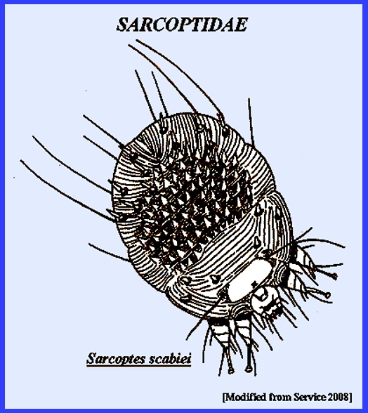
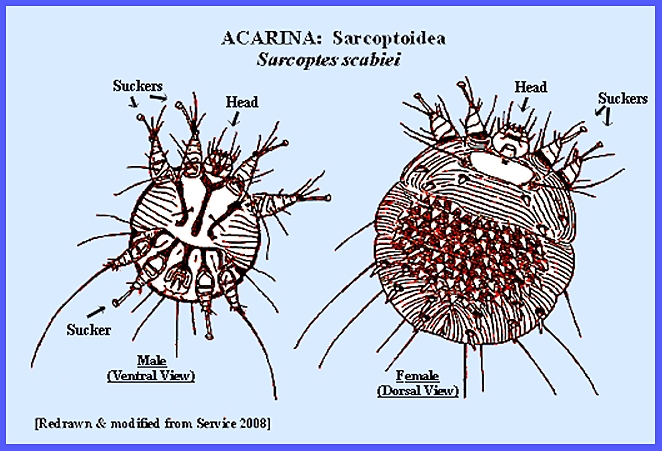
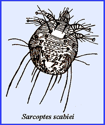
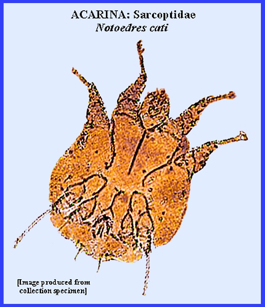
GENERAL APPEARANCE (See Photo). Females are
very tiny at barely 1/2 mm They are of pale color and rounded. There are many small spines on the dorsum
and several lines across the top and bottom of the body. The four pairs of legs are short and
cylindrical consisting of five ringed segments (Service 2008). The anterior two pairs of legs terminate
in short pedicels with suckers at the tips.
The posterior two leg pairs in females do not have suckers, ending
instead with bristles. The head is
not clearly defined, but thick short palps and chelicerae of mouthparts
extend forward from the body. LIFE CYCLE The mite
females excavate below the skin's surface where it is thin as around wrists,
fingers, feet, etc. However, most are
found on the hands and wrists and sometimes they also infest the head region
of their hosts. Once under the skin
females create winding tunnels and feed on liquids produced by the dermal
cells. About 1-3 eggs are laid daily
in the tunnels, which then require about four days to hatch with small larvae
that have only six legs. The larvae
migrate to the skin's surface where most perish, but the survivors seek out a
hair follicle where they moult and develop into a "protonymph) with
eight legs. A "tritonymph"
is produced after about 3-4 days.
Service (2008) noted that female nymphs are much larger than male
nymphs. About 3 days later the
tritonymph moults to produce either a male or female adult. Mated females then increase their size and
begin their penetration into skin.
Males, on the other hand, are much smaller and wander about the skin
surface and construct short dens for refuge.
The total life cycle usually requires less than two weeks, and females
may live up to six weeks on their hosts, but perish in a few days without
hosts. MEDICAL IMPORTANCE Skin
diseases, known as Scabies, Acariasis,
Sarcoptic Itch, etc., are
produced in humans and animals. Some
of the Sarcoptes spp.
actually inhabit tunnels underneath the skin. These mites may pass their entire lives on their hosts. Infestations among hosts are acquired by
contact. It has been estimated that
over 300 million cases of Scabies
occur annually worldwide. One family,
Sarcoptidae, and genus, Sarcoptes, is of
principal importance for humans. Sarcoptes scabiei is known as the "Human Itch Mite," of "Norwegian Itch" as it is sometimes
called. Females that are larger than
males have the dorsal part of the body marked with distinctive parallel
lines. The mites locate in the upper
layers of epidermis especially around the groin and more sequestered
areas. Mature females that bore
directly into skin where they remain concealed for a while construct egg
tunnels. Enlarging the excavation and
laying eggs follow this. Eggs hatch
in 3-4 days and the larvae leave the tunnel for the skin surface where they
enter hair follicles. Molting occurs
in 2-3 days followed by two nymphal stages.
Nymphs construct narrow tunnels where mating occurs. The life cycle varies from 8-15 days at
room temperature. Adult longevity is
3-5 weeks. A person may
acquire 50 or more mites at any given time, and any infections that develop
are not obvious for several weeks.
Following an attack there are at first few symptoms. Gradually as one becomes sensitized an
intense itching ensues, which is especially intense at night. Infections are more likely the more one
scratches the infested areas. Acquisition
of mites is through close contact with infested persons or their
clothing. Avoidance of infested areas
is preferred, but if infected one should seek medical attention from a
physician, for current products available for treatment. There are also
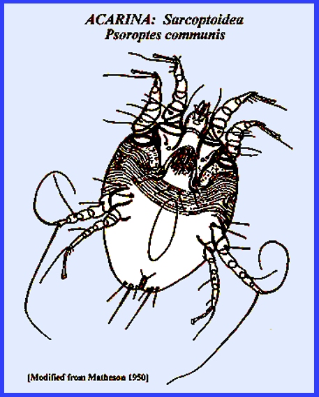
species (eg., Psoroptes communis and Notoedres cati itch mites
attacking animals that do not tunnel bur rather possess suckers for exterior
attachment to the skin. Humans only
become affected from close contact with infested animals, such as cats and
rats. CONTROL The attention
of a medical physician is advised for this group of mites, as medicinal
treatment is usually required.
Prevention involves the usual precautions of cleanliness and limiting
contact with infected surfaces, animals and people. However, Service (2008) recommended that during epidemics
clothing and bedding should be dry cleaned or washed in 50-deg. Centigrade
water. (Also See: Scabies) = = = = = = = = = = = = = = = = = = = = Key References:
<medvet.ref.htm> [Additional references may be found at:
MELVYL Library] Arlian, L. G. &
M. S. Morgan. 2000. Serum antibody to Sarcoptes scabiei and house dust mite prior to and during infestation with S. scabiei. Vet. Parasitol. 90:315–326. Arlian, L. G.,
Morgan MS, Neal J.S. 2003. Modulation
of cytokine expression in human keratinocytes and fibroblasts by extracts of
scabies mites. Am J Trop. Med Hyg. 69: 652–656 Arlian, L. G., M. S.
Morgan, & J. S. Neal. 2004. Extracts of scabies mites (Sarcoptidae: Sarcoptes scabiei) modulate cytokine expression by human peripheral blood mononuclear
cells and dendritic cells. J Med Entomol. 41: 69–73. Arlian, L. G., M. S.
Morgan, C. M. Rapp & D. L. Vyszenski-Moher. 1996. The development of protective immunity in
canine scabies. Vet. Parasitol. 62: 133–142. Arlian, L. G., C. M. Rapp, B. L. Stemmer, M. S. Morgan
& P. F. Moore. 1977.
Characterization of lymphocyte subtypes in scabietic skin lesions of naïve and sensitized dogs. Vet. Parastitol. 68: 347–358. Arlian, L. G., C. M.
Rapp, D. L. Vyszenski-Moher & M. S. Morgan. 1994. Sarcoptes scabiei: Histopathological changes associated with acquisition and expression of host
immunity to scabies. Exp. Parasitol. 78: 51–63. Kemp, D. J, S.
F. Walton, P. Harumal, & B. J. Currie .
2002. The scourge of scabies. Biologist. 49: 19–24. Matheson, R. 1950. Medical Entomology. Comstock Publ. Co, Inc. 610 p. Service, M. 2008.
Medical Entomology For Students.
Cambridge Univ. Press. 289 p Maxwell,
S. S., T. A. Stoklasek, Y. Dash, J, R., Macaluso & S. K. Wikel. 2005.
Tick modulation of the in-vitro expression of adhesion molecules by skin-derived endothelial cells. Annals Trop Med Parasitol. 99: 661–672. Orkin,
M, H. Maibach, L. C. Parish & L. M. Schwartzman. 1977. Scabies and pediculosis. Lippincott
Co., Philadelphia. p. 203. Stemmer BL, Arlian
L. G., M. S. Morgan, C. M. Rapp &
P. F. Moore. 1996. Characterization of antigen presenting
cells and T-cells in progressive scabiatic skin lesions. Vet Parasitol. 67: 247–258. |