File:
<canthariasis.htm> <Medical Index> <General Index> Site Description Glossary <Navigate to Home>
|
CANTHARIASIS (Contact) Please
CLICK on Image & underlined
links for details: "Palmer (1946) reports an
infection in a four-month-old baby, never breast-fed. The infection lasted for over four months,
the baby passing living larvae of this beetle at intervals. Infection is assumed from the feeding of
infested precooked cereals. Liggett
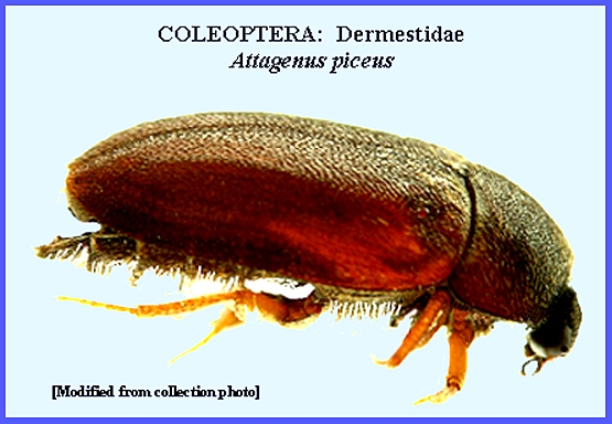
(1931) reports a peculiar rhinal myiasis in a young girl due to the invasion
of larvae of Attagenus picus Oliv. (the
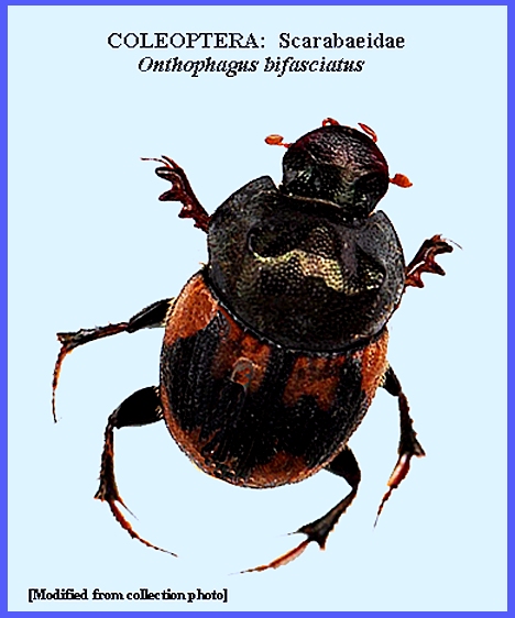
black carpet beetle). Several workers
in India, South Africa, and Ceylon have reported a peculiar type of
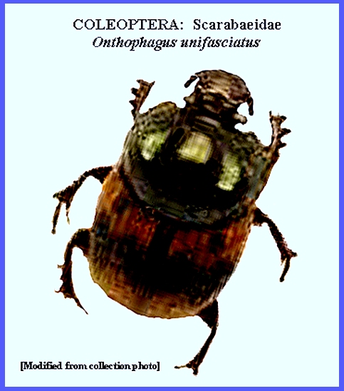
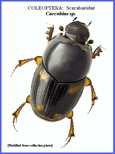
intestinal myiasis caused by the presence of scarabaeid beetles. The beetles, Onthophagus bifasciatus,
O. unifasciatus
& Caccobius mutans (see Caccobius sp.), were passed
alive in the stools. The infections
occurred only in young (three- to eight-year-old) children and the method of
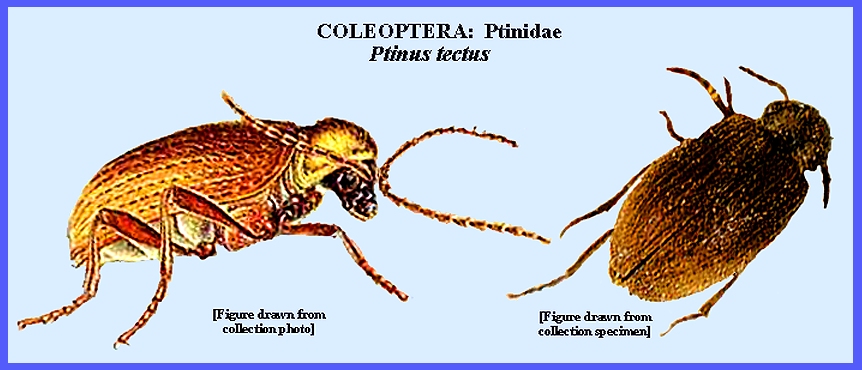
invasion may be surmised. Sharpe
(1947) lists an unusual intestinal myiasis by Ptinus tectus
(Ptinidae)." = = = = = = = = = = = = = = = = = = = = Key References: <medvet.ref.htm> <Hexapoda> Hinman, F. H. &
E. C. Faust. 1932.
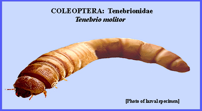
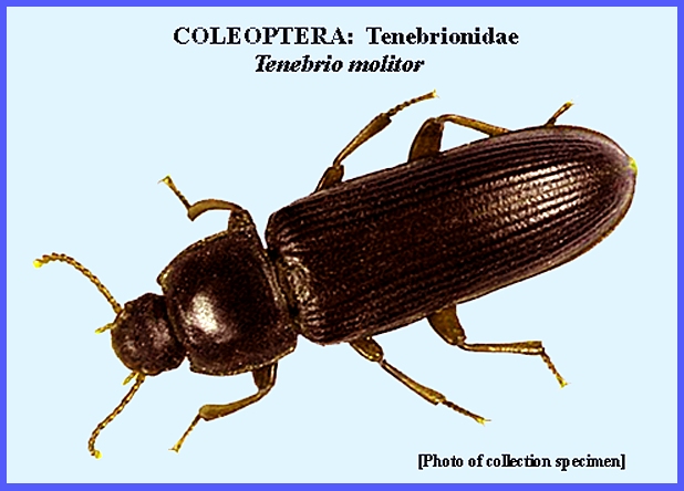
The ingestion of the larvae of Tenebrio
molitor L. (meal worm) by man.
J. Parasit. 19: 119-20. Hope, F. W.
1840. On insects and their
larvae in the human body. Trans. Ent.
Soc. London 2: 256-271. Legner, E. F. 1995. Biological
control of Diptera of medical and veterinary importance. J. Vector Ecology 20(1): 59_120. Legner,
E. F.. 2000. Biological control of aquatic
Diptera. p. 847_870. Contributions to a Manual of Palaearctic
Diptera, Vol. 1, Sci. Herald,
Budapest. 978 p. Liggett, H.
1931. Parasitic invasion of
the nose. J. Amer. Med. Assoc.
96: 1571-72. Matheson, R. 1950. Medical Entomology.
Comstock Publ. Co, Inc. 610 p. Palmer, E.
D. 1946. Intestinal canthariasis due to Tenebrio
molitor. J. Parasitol.
32: 54-55. Service, M. 2008. Medical
Entomology For Students. Cambridge
Univ. Press. 289 p Sharpe, D. S.
1947. An unusual case of
intestinal myiasis. British Med. J.
1: 54. ADDENDUM Xi Sun, L-Fu Wang et
al. 2016. A case report: A rare case of infant
gastrointestinal canthariasis caused by larvae of Lasioderma
serricorne (Fabricius, 1792) (Coleoptera:
Anobiidae). Infect. Disease Poverty
5: 34. A case of an eight-month-old baby girl with irritable
feeling, rubbing eyes, history of contact with mud and eating oranges twice
during five days before attendance, and having “worms” in her stool was
admitted to the First Affiliated Hospital of Sun Yat-sen University,
Guangzhou, China. The clinical examination revealed that the pulse rate,
blood pressure and temperature were regular, and the examination of the head,
neck, and chest were unremarkable. The stool specimens containing “worms”
were sent to the Department of Parasitology, Zhongshan School of Medicine,
Sun Yat-sen University. The worms were recovered, studied morphologically
using naked eyes and anatomical lens, PCR analyzed targeting cytochrome
oxidase subunit 1 (COX1) and 18S rRNA genes,
examined by sequence analyses of the PCR products and finally classified by
phylogenetic analysis to identify their species. Based on the findings, the
worms were diagnosed as the larvae of L. serricorne. |