File: <arachnidamed.htm> <Medical Index> <General Index> Site Description Glossary <Navigate
to Home>
|
Arthropoda:
ARACHNIDA Ticks, Mites
Spiders Pseudoscorpions (Contact) Please CLICK on Images to enlarge & underlined links for details: GENERAL
CHARACTERISTICS OF ARACHNIDA
The general
characteristics are the absence of antennae and a body comprised of a cephalothorax and an abdomen, the
latter may appear as only a single part without divisions. The cephalothorax
bears four pair of walking legs and 6-8 eyes raised on tubercules. The head
appendages include chelicerae,
which are jaw like with claws and poison
duct openings at their tips.
The basal portion of pedipalps
serves both feeding and sensory functions. The Arachnida
are air-breathing arthropods, the body of which is divided into two
parts: (1) The cephalothorax,
including the fused head and thorax, and (2) The abdomen. The abdomen can be
either segmented or unsegmented. The mites and ticks have their entire body
fused to form forms a sac. The head appendages are highly modified. The antennae are lateral and the eyes,
when they occur, are simple and sessile, and the eyes, when present, are
rather simple and sessile. In the adults there are four pairs of ambulatory
legs that are attached to the cephalothorax. The first developmental stage is
the larva, which has three pairs of legs.
When respiratory organs occur they are either book lungs or
tracheae. The sexes differ
structurally and metamorphosis is incomplete. The immatures resemble small adults. The arachnids imbibe fluid from their prey by means of a
"sucking stomach."Their mouthparts function either for crushing
their prey and sucking up the liquid portions or for piercing and cutting the
host tissues to obtain blood (Matheson 1950). The
mouthparts consist of a pair of chelicerae located in front of the mouth
opening; a pair of pedipalpi that are located on the mouth sides or just
posterior to them. In some parasitic
species there is a structure called the "hypostome" that is located
directly beneath the mouth opening.
Chelicerae vary structurally in different orders. In the spiders (Araneida) each chelicera
consists of a large basal segment and a terminal one shaped into a claw. Spiders used these structures to capture
and kill their prey. A poison gland
is located near the tip of the claw.
Parasitic species (e.g., ticks) use the chelicera as piercing and
cutting tools. The pedipalpi resemble legs in all the groups have 4-6
segments. In the spiders the pedipalpi 4 of the male are greatly modified
into very specialized organs insemination of females. Among many of the ticks
they are protect the highly developed piercing organs (Matheson 1950). The following
descriptions include both groups of medical and non-medical importance for
distinction purposes: - - - - - - - - - - - - - - - - - - - - - - - - - - - - - -
- - - - - - - - - - - - - - - - - - - - The Order Araneae -- includes the true
spiders. Segmentation is obscure in
the abdomen and there are no obvious appendages except 3-4 pairs of
spinnerets at the posterior end of the abdomen that are modified abdominal
appendages. Several examples of
spiders may be seen in the following diagrams Inv143 - Inv147: Food & Digestion -- Insects and
other small animals are caught in webs.
The prey is paralyzed and their liquid contents are moved up through
the pharynx and esophagus. A sucking stomach pumps food from the
prey through the mouth and into the digestive tract. Nine
diverticulae from the intestine lead to various body parts. There is one located forward and four on
each side, which function to increase the surface area. The posterior part of the intestine is
surrounded by digestive glands and some food may actually enter the
glands. A rectal caecum occurs at the junction of the rectum and
intestine. Circulation -- The heart is long and
located in the abdomen. The dorsal aorta in the cephalothorax
has subsequent branches to appendages and the brain and eye regions. Some blood is pumped posteriorly to a
short posterior aorta. The haemocoel is divided into various
sinuses. Blood reaches the book lungs
and is aerated after which it returns to the heart. Respiration -- Air diffuses directly
into the book lungs, as the
blood does not carry oxygen. Some
tracheae may occur but they are never well developed. Excretion -- Malpighian tubules
serve for excretion. Coxal glands that are modified
nephridia may also be involved in excretion. Nervous System -- There is a typical
pattern where a great concentration of ganglia occurs in the anterior
cephalothorax. Nerves run out to
different parts of the body. Sensory Organs -- There are the
eyes, pedipalps and setae all over the body all of which have sensory
functions. Reproduction -- The sexes are
separate. Ducts open near the
anterior end of the body, but fertilization is internal. Males use
pedipalps to transfer sperm from their genital pore to that of the
female. Eggs are laid in silken
cocoons and maternal care is common.
Development is direct. Silk
Glands -- There are several varieties of silk glands. The silk they produce differs in strength,
slipperiness, etc. Different kinds of
webbing are produced for particular circumstances. The tips of the legs are modified for walking on the webs. Economic Importance -- Some species
of spiders are poisonous to humans and animals. Spider silk has been used in bombsights during World War II. ------------------------------------ Order: Scorpiones
(Scorpionida) -- scorpions: These
animals have a well marked cephalothorax and segmented abdomen that is
equipped with a sting and poison gland at the posterior end. They can be dangerous in warmer
regions. Chelicerae and pedipalps are
both chelate. They have book
lungs. They feed on other
arthropods. They are also viviparous
as they bear living young. See Inv150 & Inv151 for examples: ------------------------------------ Order:
Amblypygi. (Pedipalpia) -- whip spiders and tailless whip scorpions: There is a long tail, large palps and
small chelicerae. ------------------------------------ Order:
Pseudoscorpionida -- book scorpions:
These are small animals that have the appearance of scorpions because
their pedipalps are pincers. The
abdomen is rounded but without a sting.
They feed on small insects. See Inv152 for example: ------------------------------------ Order:
Opiliones (Phalangida) -- harvestmen: Their
extremely long walking legs have earned them the name of "Daddy Long Legs." The body regions are all compacted into a
single division. They are predators
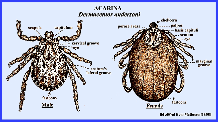
of small insects and other arachnids. See Inv154 for example: ------------------------------------ Order: Acarina -- mites and ticks:
The chelicerae and pedipalps are modified into projections called a hypostome. They are parasites and vectors of disease, and serious pests of
vegetable and tree crops. See Inv153 for example: ------------------------------------ Class Pycnogonida -- sea spiders: These are tiny marine animals. Included are parasites, commensals and
free-living predators. ------------------------------------ Class: Merostomata: Order: Xiphosura -- horseshoe crab:
The range is from the East Coast of North America to the coasts of
southeastern Asia. These animals have
remained essentially unchanged sinde the Paleozoic. They and the Pycnogonida are the only marine arachnids. They are also the only Arachnida with
compound eyes. The chelicerae are
chelate and the pedipalps look like walking legs. But there is four pair of true walking legs. The abdomen has well developed appendages
that have been modified into book gills. Horseshoe
crabs are of course a misnomer as they are not mollusks. Their blood, which is blue in color, is high
in metallic copper and is harvested regularly for medical research. See Inv148 & Inv149 for examples: Subphylum: Myriapoda,
Class: Chilopoda includes the centipedes.
They are dorso-ventrally flattened.
Their body consists of a head and trunk but there is no thorax nor
abdomen. The head bears one pair of
antennae, one pair of mandibles, one pair of maxillipedes with poison
glands at the bases and ducts leading to pointed tips (Note: these are absent in the Diplopoda). There are two pairs of simple eyes called pseudocompound eyes. They have maxillae on the 1st and 2nd
segments. The trunk bears uniramous
appendages and there are 15 to 175 segments.
See examples at Inv141. Body
Wall -- This consists of a cuticle, muscles and a haemocoel Digestive Tract -- A typical mouth
to anus arrangement. Circulatory System -- The heart is
tubular with one pair of ostia per segment.
The blood does not
carry oxygen Respiration -- The tracheae are
lined with ectoderm and cuticle, and heavy rings of cuticle line them. They branch out and ultimately reach all
tissues of the body. The blood does
not have an oxygen carrying function. Excretion -- Malpighian tubules are long, thread-like and blind-ending
tubules. They lie in the haemocoel
and empty into the digestive tract at the junction of the mid and
hindguts. They extract nitrogenous
wastes from the blood. Nervous System -- This system is the
same as that found in the Crustacea. Reproduction -- The sexes are
separate. Genital organs are found at
the posterior end of the body and development is direct. Locomotion -- These animals are fast
movers. Long posterior legs are
sensory and used when moving backwards. Food & Digestion -- Chilopoda
are carnivorous and their food is paralyzed first by the maxillipedes. MEDICAL
IMPORTANCE OF THE ARACHNIDA The Arachnida
are divided into about nine orders with six of these being primarily of
medical importance (Matheson 1950).
One group, the Acarina, is most encountered (See: Tick Borne Diseases). The other five orders do contain species
that have poison glands, and their bites or stings can be of such severity as
to require medical attention. Some species are vectors of pathogenic agents
(Matheson 1950) and Medical Entomology. Table 1. Tick Species That Inflict Harmful Bites
|
|||||||||||||||||||||||||||||||||||||||||||||
|
= = = = = = = = = = = = = = = = = = = = = Key References: <medvet.ref.htm> [Additional references may be found at:
MELVYL Library] Brumpt, E. 1927. Précis de
paraaitologie. 4th ed. Paris, France. Davis, G. E. 1942. Tick vectors and
life cycles of ticks. IN: Symposium on relasping fever in the
Americas. Amer. Assoc. Adv. Sci. Pub. 18:
67-76 Dunlop, J. A. &
M. Webster. 1999. Fossil evidence, terrestrialization and arachnid phylogeny.
J. Arachnol. 27: 86-93. Harvey, M. S. 2002.
The neglected cousins: What do we know about the smaller Arachnid
orders? Journal of Arachnology 30(2): 357-372. Harvey, M. S. 2007.
The smaller arachnid orders: diversity, descriptions and distributions
from Linnaeus (1758 to 2007). Pages 363-380 in: Zhang, Z. Q. & W. A.Shear
(eds.) Linnaeus Tercentenary: Progress in Invertebrate Taxonomy.
Zootaxa 1668: 1–766. Harvey, Mark S. 2002.
The neglected cousins: what do we know about the smaller arachnid
orders?. J. Arachnol. 30(2): 357-372. Mail, G. A. & J.
D. Gregson. 1938. Tick paralysis in British Columbia. J. Canad. Med. Assoc. 39: 532-537. Matheson, R. 1950. Medical Entomology. Comstock Publ. Co, Inc. 610 p. Nuttall, G. H. F.
1908. The Ixodoidea or ticks,
spirochaetosis in man and animals, piroplasmosis. Harben Lectures. J. Roy
Inst. Pub. Hlth, July, Aug, Sept. Patton, W. S. & F.
W. Cragg. 1913. A textbook of medical entomology. Calcutta & London. Patton, W. S. & A. M. Evans. 1929-1931. Insects, ticks, mies and venomous animals of medical and
veterinary importance. Part I. Medical; Part 2, Public Health. Croydon, England. Service, M. 2008.
Medical Entomology For Students.
Cambridge Univ. Press. 289 p Shultz, J. W. 1989.
Morphology of locomotor appendages in Arachnida - evolutionary trends
and phylogenetic implications. J. Linn. Soc. 97: 1-56. Shultz, J. W. 1990. Evolutionary morphology and
phylogeny of Arachnida. Cladistics 6: 1-38. Shultz, J. W. 1994. The limits of stratigraphic evidence
in assessing phylogenetic hypotheses of recent arachnids. J. Arachnol. 22:
169-172. Shultz, J. W. 2007.
A phylogenetic analysis of the arachnid orders based on morphological
characters. Zoo. J. Linn. Soc. Zoological 150(2): 221–265. Starobogatov, Y. I. 1990.
System and phylogeny of Arachnida (analysis of morphology of paleozoic
groups) [Russian]. Paleontologicheskii Zhurnal 24: 4-17. Weygoldt, P. & H. F. Paulus. 1979.
Untersuchungen zur Morphologie, Taxonomie und Phylogenie der
Chelicerata. 1. Morphologische
Untersuchungen.. Zeit. für Zool. Syst. u. Evolutionsforschung 17:
85-116. Weygoldt, P. 1998.
Evolution and systematics of the Chelicerata. Exptal. & Appl.
Acarol. 22: 63-79. |


