File: <sarcophagidaemed.htm> <Medical Index> <General Index> Site Description Glossary <Navigate to Home>
|
SARCOPHAGIDAE (Flesh
Flies) (Contact) Please
CLICK on Images
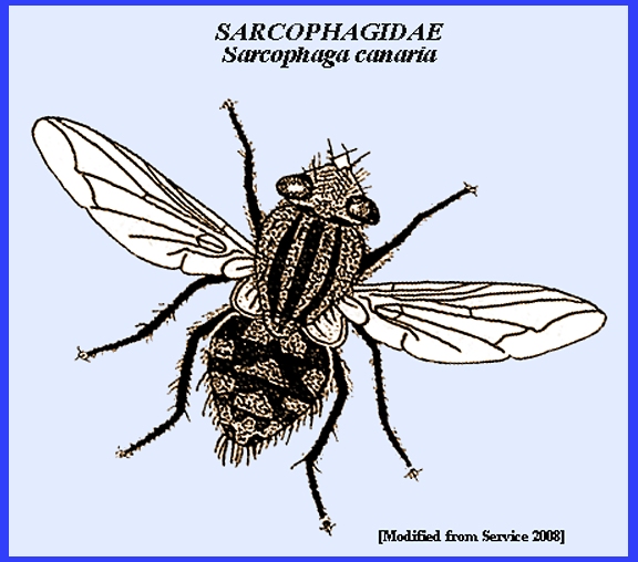
& underlined links to view: Sarcophagidae. -- <Habits>; <Adults> & <Juveniles> -- The flesh flies
are similar to Calliphoridae, but they are usually black or gray with stripes
on their thorax. Adults feed on sweet
foods such as flower nectar, fruit juice and honeydew. Their
larvae show diverse habits, but most feed on animal material, with many being
scavengers. Some species are
scavengers, some are parasites of other insects and a few are parasites of
vertebrates that develop in skin wounds. Description
& Statistics
Early reviews of host preferences were by Aldrich (1915)
and Greene (1925a). Entomophagous
species are in subfamilies Sarcophaginae and Melanophorinae. Many species of Sarcophagidae are limited
to carrion, others to manure; but there are both predaceous and parasitic
species. Predaceous species attack
egg pods of Acrididae. Well-known
genera having this habit are Sarcophaga
and Blaesoxipha. Oophagomyia
and Wohlfahrtia are predaceous
in the egg capsules of the same host group, and an occasional species of Blaesoxipha is both parasitic on the
active stages and predaceous on the eggs.
Mantidophaga is an
internal parasitoid of the late nymphs and adults of Mantidae. Clausen (1940) noted that an unusual degree of plasticity
was revealed in the behavior of species of this family, and many were
apparently only in the transitional stages to obligate parasitism. Most parasitic species are primary,
solitary, endoparasitoids, though gregarious species are known. The principal host range includes
Orthoptera (Acrididae, Mantispidae) and Lepidoptera, but other insect orders
may be attacked. A few species are
parasites of snails, while others are carrion feeders or vertebrate parasites. A host group frequently attacked by species of the
Sarcophaginae is the social wasps and bees.
The relationship ins some cases is strictly parasitic, while in others
it is commensal. Myiapis and Senotainia are internal parasitoids of worker honeybees,
and Sphixapate develops within
the larvae. Metopia and Brachicoma are external parasitoids or predators of the
brood of wild bees, the latter genus attacking mainly bumblebees. Hilarella
and Miltogramma develop on
various insects, which are stored in cells of hunting wasps, or on the
material with which the cell of bees are provisioned. Several genera have widely different host
preferences. Lepidopterous larvae and
pupae frequently yield sarcophagid flies, and it has been thought that there
were parasitic. Several species of Sarcophaga associated with the gypsy
moth were found to be scavengers only (Patterson 1911). Young larvae were unable to enter healthy
larvae or pupae, and if artificially introduced into the bodies of living
individuals they died. However,
species of Agria are predaceous
on pupae of Lepidoptera. Eleodiomyia has been reared from adult
beetles of the family Tenebrionidae; Scarabaeophaga
from pupae and adults of Cotinus
nitida L.; and Sarcophaga spp. from adult Pentatomidae,
Blattidae, etc (Clausen 1940/62). Arachnidomyia sp. has been reared from
egg sacs of spiders and various genera and species from snails. There is a wide range in host preference found among the
parasitic and predaceous species of Sarcophaga. Not much is known regarding insect hosts
of Melanophorinae, although species have been reared occasionally from spider
egg masses and from coleopterous larvae and adults. Melanophora, Cirillia, and closely related forms are
parasitic in Isopoda (Porcellio,
Metaponorthus, Oniscus), and some species of this
subfamily have been reared from snails.
Biology & Behavior
In Brachycoma
lineata, S. lellyi
and S. caridei, the maggots enter the host body
through the thin membrane at the base of the wing. S. filipjevi enters through the membranes
of the abdomen or through the genital opening. The latter behavior is similar to that of Eleodiomyia in attacking tenebrionid
beetles. Wood (1933) noticed that the
maggots of S. destructor readily enter freshly molted
hosts but are not able to if the integument is fully hardened. The host dies within a short time after
the larvae have entered the body.
Mature larvae of S. linerata and S. caridei
emerge from the host while the latter is still alive, and some parasitized
individuals may recover. However, the
hosts of Wohlfahrtia are
usually dead before the larvae finish feeding. They usually emerge through the thin membranes of the neck,
although some individuals of S.
kellyi are believed to emerge
through the anal opening. Wood (1933) found that 78 % of attacked hosts of Brachycoma lineata recovered, but only
38% were able to reproduce thereafter.
Relatively little growth occurs in S.
destructor as long as the host
remains alive. The young larvae of
this species attack the wing muscles, nd death results primarily through
infection. After this, development of
the parasitoid is rapid. Only 16% of
hosts containing one parasitoid larva died, while 92% died when two or more
were present. If hosts are immature
at the time of attack, they do not attain the adult stage. Larval feeding is confined mostly to the
fat body. The number of individuals
developing in each host varies, being usually only 2 in the case of Brachycoma lineata, a maximum of 11 in B. filipjevi and 9 in B.
caridei. There is often a high percent parasitization by
Sarcophagidae, but opinions vary as to their value in natural control. Smith (1915) stated that swarms of Dissosteira longipennis Thoms. in New Mexico were almost eliminated by
S. kellyi. Kunckel d'Herculais
(1894) found parasitization of Schistocerca
by sarcophagids in Algeria to be 69% in 1889 and 75% in 1890. The flies followed host swarms, harassing
them continuously. In the case of Wohlfahrtia euvittata in South Africa, 50-90% of Locustana were found parasitized, and in
some areas this attack was responsible for discontinuing poisoning programs. Some species that were discussed as internal parasitoids of
nymphs and adults of locusts are also predaceous in egg masses of the same
hosts. This range in habit has been
found for Sarcophaga opifera Coq. in British Columbia, and
Treherne & Buckell (Clausen 1940/62) thought that the larvae, after
leaving the body of the adult host, continued their development on the eggs
in soil. Potgieter (1929) in South
Africa observed that W. euvittata is very important in natural
control of Locusta pardalina Wlk., when parasitic on the
active stages. About 50% of the egg
masses in one area were destroyed by this fly. The maggots are laid in groups in the openings of partly
hatched egg pods or in the froth at the upper ends of those freshly laid or
exposed. Larvae in various stages of
development were found on the surface of the ground, and these were migrating
to other egg pods for further feeding (Potgieter 1929). Sarcophagids that are parasitic or predaceous on the brood
of bees and wasps are mostly in genera Metopia,
Brachicoma and Hilarella. Bougy (1935) described the attack of H. stictica
Meig on Ammophila hirsuta Scop. in France. The host stores its nest with noctuid
larvae, and the female fly appears while the prey is being transported to the
nest. She does not attempt to
larviposit on it at this time. It is
only after the caterpillar has been placed in the cell and the Ammophila egg laid that she evades the
host, enters the burrow and lays her own minute larva alongside the host
egg. This egg is consumed within 24
hrs., and the larva then enters the body of the caterpillar to complete its
development. Each individual may be
regarded as a predator on the egg of Ammophila
and an internal parasitoid of noctuid caterpillars. Mature larvae and young pupae of bumblebees are parasitized
by Brachycoma sarcophagina Tns. in North America. The live young are laid on or in the brood
cells. They enter the body and feed
until larval maturity. Pupation
occurs in the nest material at the bottom of the comb. B.
davidsoni Coq. is thought to
lay eggs directly on the larvae; and after one is consumed, the parasitoid
larva enters other cells to attack their occupants. Metopia leucocephala Rossi has been found in
cells of Philanthus. Females enter the host burrow for a short
distance and there lay their larvae, which have to find their own way to the
cells, sometimes several feet away.
Adult honeybees are found heavily parasitized by Senotainia tricuspis Meig. in some parts of Russia. The larvae feed principally in the
thoracic region, the same habit being recorded for Myiapis angellozi
Seguy (Seguy 1930). Agria mamillata
Pand. is predaceous on pupae of Hyponomenta
in Italy, with flies appearing in the field in early June to lay their
partially incubated eggs on caterpillars when they are mature but before
cocoon formation. The young larva
enters the body of the pupa and quickly consumes its contents. It then penetrates the adjoining cocoons
and continues its feeding, destroying 50 or more pupae per single larva
before maturity (Servadei 1931). S. latisterna
Perk was reared from various pupae of Lepidoptera, where it was believed to
be a true facultative parasite (Hallock 1929). Thompson (1920a, 1934) studied several sarcophagids that
are parasitic in isopods of genera Oniscus,
Porcellio and Metaponorthus. They differ in several ways from the general habits of the
family. The adaptive characters of
the 1st instar larvae, as well as the habits of the immature stages, show a
closer biological affinity with Tachinidae than by any other members of
Sarcophagidae. Parafeburia maculata Fall, is a solitary internal parasitoid of the
first two genera. Its unincubated
eggs are probably laid in the general vicinity of the hosts or where they are
in the habit of congregating. They
hatch in ca. one week when these membranous eggs give rise to planidium type
larvae. This is the only instance in
the Muscoidea in which this larval form hatches from membranous eggs that are
unincubated at the time of laying.
Young larvae enter the host body through the soft cuticle separating
the ventral sclerites or at the bases of the appendages. Once inside the host, the larva is found
with its posterior end fixed in a perforation in the integument, and a
respiratory funnel is formed. The 2nd
instar larva has a very thin integument, and tests have shown that an
exchange of gases takes place through it;
The greater part of the oxygen requirements of the larva may be
secured in this way, and pupation occurs within the remains of the host. Clausen (1940) commented on the definite effect on the
reproductive system and the secondary sexual characters of the host as a
result of parasitism by Parafeburia. Female ovaries are atrophied, owing to
absorption of fat by the parasitoid, and such females do not develop a brood
pouch. Less complete information
regarding Cirillia angustifrons Rond. was presented by
Thompson (1920a). General habits are
similar to those for Parafeburia,
with the outstanding distinction in the host relationships being the
formation of the integumentary respiratory funnel by the larvae. This habit is unknown elsewhere in
Sarcophagidae, although it is common in Tachinidae, indicating a higher
development of the parasitic relationship than has been attained by other
species. Life
Cycle
The life cycle of parasitic Sarcophagidae, from
larviposition to adult emergence, is relatively short, being completed in
16-30 days, of which the larval feeding period takes only 5-10 days. In Wohlfahrtia,
larval maturity is followed by a resting period of 6-12 days, and the pupal
stage then requires 16-30 days.
Several generations are usually produced each year, and 5-6 are
recorded for S. kellyi.
Hibernation is known for Brachycoma
lineata and S. kellyi, in both of which mature larvae
rather than pupae persist through winter.
The life cycle of P. maculata in Oniscus and Porcellio
differs from the general habit of the family by having only a single
generation each year. Adults appear
in midsummer, and winter is passed as 2nd instar larvae within the live host
(Clausen 1940/62). For detailed descriptions of immature stages of Sarcophagidae,
please see Clausen (1940/62). = = = = = = = = = = = = = = = = = = = = Key References: <medvet.ref.htm> <biology.ref.htm>, [Additional references
may be found at: MELVYL Library] Allen, H. W. 1926. Proc. U. S. Natl. Mus. 68:
1-106. Clausen, C. P. 1940/1962. Entomophagous Insects.
McGraw-Hill Book Co., Inc., NY. & London. 688 p.
[reprinted 1962 by Hafner Publ. Co.]. Cole, F. R. 1969. The
Flies of Western North America. Univ.
Calif. Press, Berkeley & Los Angeles.
693 p. Downes, W. L.,
Jr. 1965. Family Sarcophagidae IN:
A. Stone et al. A catalog of the Diptera of America north of Mexico
United States Department of Agriculture, Washington,
D.C. Hertig, M. 1942.
Phlebotomus and
Carrion's disease. Amer. J. Trop.
Med. 22: Suppl. Hertig, M. and G. B.
Fairchild. 1948. The control of Phlebotomus in Peru with
DDT. Amer. J. Trop. Med. 28: 207-30. James, M. T. & R. F.
Harwood. 1969. Herm's Medical Entomology, 6th ed. MacMillan Co. 484 p. Kano, R., Field, G.
& Shinonaga, S. 1967. Fauna Japonica: Sarcophagidae (Insecta:
Diptera) Biogeographical Soc. Japan. Distributor: Tokyo Electrical Engineering College Press. Kunckel D'Herculais,
J. 1894.
Les Dipteres parasites des Acridiens: les Muscidae vivipares a larves
Sarcophages. Aptenie et castration parasitaire. Acad. Sci. Compt. Rend.,
Paris 118: 1106. Legner, E. F. 1995. Biological control of Diptera of medical and veterinary
importance. J. Vector Ecology 20(1):
59-120. Legner, E. F.. 2000.
Biological control of aquatic Diptera. p. 847-870. Contributions
to a Manual of Palaearctic Diptera, Vol. 1, Science Herald, Budapest. 978 p. Lehrer, A.Z. 2006.
Sarcophaginae et Paramacronychiinae du Proche Orient(Insecta, Diptera,
Sarcophagidae). Pensoft Series Faunistica 60: Pensoft Publishers, Sofia-Moscow. pp.
165-240. Lehrer, A.Z. 2003.
Sarcophaginae de l'Afrique (Insecta, Diptera, Sarcophagidae) In:
Entomologica, Bari, 37: 5-528. Lehrer, A.Z. 2010. Taxonomic Atlas of the postabdominal
structures Sarcophagidae (Insecta, Diptera). Vol. 1 In: Entomologica, Bari,
42: 3-459. Matheson, R. 1950. Medical Entomology. Comstock Publ. Co, Inc. 610 p. Pape, T. 1987.
The Sarcophagidae (Diptera) of Fennoscandia and Denmark. Fauna Ent. Scandinavica. 203 pp. Pape, T. 1998.
Sarcophagidae.-pp. 649–678, IN:
Papp, L. & Darvas, B. (eds), Contributions to a manual of
Palaearctic/European Diptera. Science Herald; Budapest. Pape, T. 1996.
Catalogue of the Sarcophagidae of the world (Insecta: Diptera).Memoirs of
Entomology International 8: 1-558 Potgieter, J.
T. 1929. A contribution to the biology of the brown swarm locust Locustana pardalina (Wlk.) and its natural
enemies. Proc. Agr., Union So. Afr., Dept. Agr. & For.,
Pan-Afr. Agr. Vet. Conf., Pretoria.
p. 265-308. Roback, S. S. 1954.
Illinois Biol. Mon. 23: 1-181. Rohdendorf, B. B. 1988.
Family Sarcophagidae in Bei-Bienko, G. Ya. Keys to the Insects
of the European part of the USSR Fauna SSR (NS) 12: xv, 1-496. [In Russian; English
translation 1988, pp. 1021–1096; Washington, D.C.] Rohdendorf, B.
B. 1930-1975. Sarcophaginae,in Lindner, E. Fliegen die
Palaearktischen Region. 11 64h, 1-232;1985, 64h. Sarcophaginae (Lieferung 330) 1- 297;1993 64h.
Sarcophaginae (Lieferung 331) 1-441, 90 Abbildungen (figures). Venturi, F., 1960. Sistematica e geonemia
dei Sarcofagidi (escl. Sarcophaga s.l.) italiani (Diptera). Frustula Entomologica, 2 (7): 1-124. Service, M. 2008.
Medical Entomology For Students.
Cambridge Univ. Press. 289 p Verves, Yu.G. 1986. Family Sarcophagidae. In: Soós Á.
& Papp L. (eds.), Catalogue of Palaearctic Diptera, 12. Akadémiai Kiadó,
Budapest Elsevier, Amsterdam pp. 58-193. Wood, O. H. 1933.
Notes on some dipterous parasites of Schistocerca and Locusta
in the Sudan. Bull. Ent. Res.
24: 521-30. |

