:
<bacteria.htm> <Index to Mycology> Pooled References <Glossary> Site
Description <Navigate to
Home>
The Bacteria: Monera Schizomycophyta1
(Contact) Please CLICK on underlined
links & included illustrations for details Use Ctrl/F to search
for Subject Matter: This group of organisms is discussed here because of
the Actinobacteria (Actinomycetales), which have been considered as an
intermediate group between fungi and bacteria. Bacteria have traditionally been difficult to classify because
of their ability to mutate rapidly and their morphological similarities. Biochemical instead of morphological means
of identification has been deployed more successfully. The Class
Schizomycetes,
Order Eubacteriales
are
the true bacteria. They are non-filamentous,
non-photosynthetic structures with heavy cell walls. They include the bacilli, cocci and
spirilla. [See PLATE 4 for flagella arrangements] Clamydobacteriales are the sheath
bacteria. They possess a common sheath, which holds
individual bacterial cells, thereby approaching a filamentous form. The sheath is composed of Fe(OH)3
and Mn(OH)3 Spirochaetales have
long cells. Myxobacteriales are
the slime bacteria. In class rods are distributed in a common,
mucilaginous mass. Although they are
individual organisms, the whole mass behaves as a unit. The mass (sheet) concentrates in one area,
assumes a stalk shape and then branches in some species. Actinomycetales are the ray bacteria. They appear to bridge the gap between
bacteria and true fungi. In acid
soils the population density is relatively low, as the optimum pH for growth
is between pH 7 & 8. Some genera,
e.g., Streptomyces and Actinomyces, are able to decompose many
substances. Phages have been known to attack
this group, but no phages are known for the fungi. The mycelium ranges between 0.7 and 1.0 microns in diameter. Characteristics that make the
Actinomyctales similar to fungi are a branching aerial mycelium, the
production of conidia in some species and motile spores. They are similar to bacteria in the
diameter of their spores and filaments, many reproduce by rods, there is an
absence of sexual reproduction, a flagella may be present, they are attacked
by phages and they are not effectively attacked by antibiotics. In the family Mycobacteriaceae the
mycelium is rudimentary or absent. In
these acidfast organisms there is a tendency toward filament formation and
branching. A representative Genus is Mycobacterium. Some diseases caused by members of this
family are leprosy
and tuberculosis. The family Actinomycetaceae has
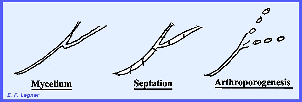
a mycelium that septates, and individual cells break away to form Arthrospores
that are aerial and possess no conidia. The Genus Actinomyces is
anerobic or microaerophilic, parasitic and not acidfast, while the Genus Nocardia
is aerobic, partially acid fast or non-acid fast. Two species of Actinomyces act as parasites on animals
causing Actinomycoses
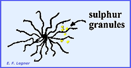
(lumpy jaw). The mycelium radiates from the site of
infection in a characteristic manner.
The species develop anaerobically.
[See PLATE
5 for life cycle]. The genus Nocardia, with over
43 species, is aerobic, has an aerial mycelium that breaks up early in the
developmental cycle and uses paraffin as a carbon source. The disease Madura Foot is associated with
this group. In the Streptomycetaceae an
aerial mycelium is common and conidia are formed. The Genus Streptomyces has over 150 species, thirty
percent of which are used for antibiotics production.
This is different from the Actinomyces where no antibiotics are
produced. Streptomyces plays
an important role in the ecology of microflora. Streptomyces scabies (has been referred to as Actinomyces
scabiea in the early literature) attacks potato causing the common potato scab
disease. It may be
controlled in acid soil. The
non-septate mycelium shows profuse branching.. Apices may form spirals on the conidiopores that then form
septae and finally break-up to form rod-like conidia or catenulate
conidia. [See PLATE 5 for life cycle
and PLATE 6
for examples of several
species]]. In the Genus Micromonospora, which is widely
distributed in lakes and in lake mud, there is a bulb-like cell at the top of
the conidiophore. ------------------------------------------- Please see PLATE
6 for
examples of Actinomycetales cultures on agar and the following for additional structures: Plate 4 = Flagella Arrangement --
Eubacteriales
Plate 5 = Life Cycles –
Actinomycetales Plate 64 =
Bacterial cell arrangements: Micrococcus,
Diplococcus, Streptococcus, Staphylococcus, Sarcina,
Microbacillus, Diplobacillus, Streptobacillus, Microspirillum
& Diplospirillum. Plate 65 =
Capsulated cells of bacteria. Plate 66 =
Flagella arrangements: Monotrichous,
Lophotrichous, Amphitrichous & Peritrichous. Plate 67 =
Life Cycle -- Streptomyces sp. |


