File:
<internalanatomy.htm> < (Entomology),
(Invertebrates), (General
Index)> <Invertebrate Bibliography> <Glossary> <Site Description>
< Home>
|
Entomology: INTERNAL
ANATOMY 1 Kingdom: Animalia, Phylum: Arthropoda Subphylum: Hexapoda: Class: Insecta: Entomology Internal
Anatomy (Contact) Please CLICK on underlined
categories to view and on included illustrations to enlarge: Depress Ctrl/F to search for subject matter: |
|
Insecta:
Entomology Internal Anatomy |
|
|
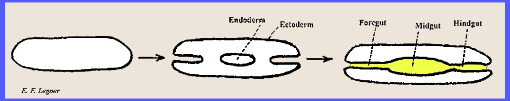
Embryological Development.
-- During embryological development the material that lines the hind and fore
guts is the same as the integument that forms the insect body. It is usually called cuticula also frequently intima.
When molting, the cuticula is cast
off including that which occurs in the fore and hind guts. The principal absorptive area for
digestible materials occurs in the midgut.
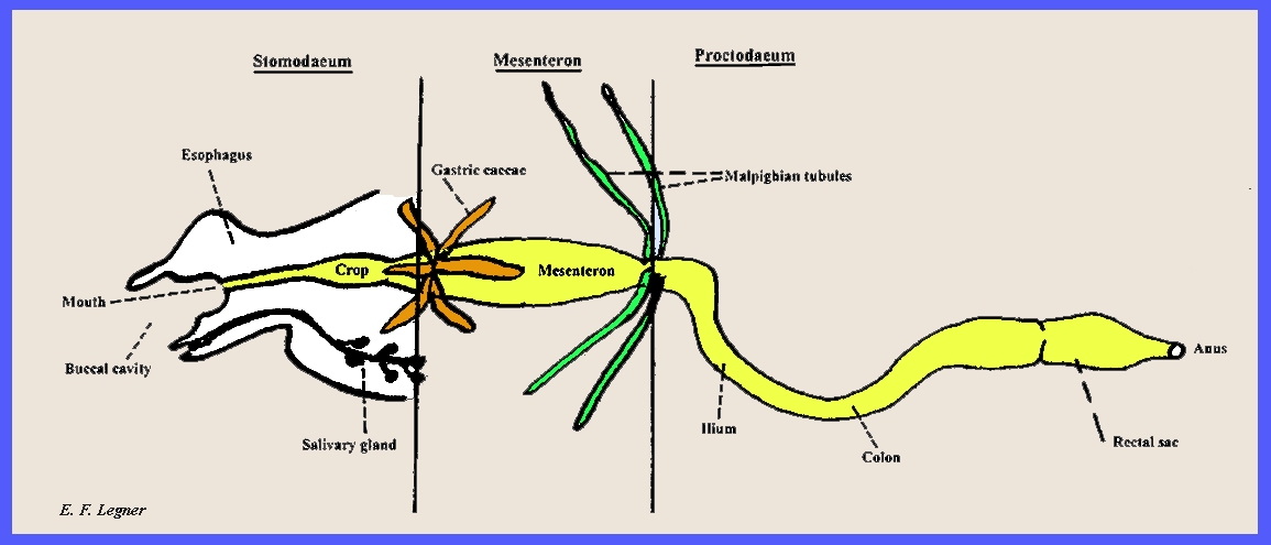
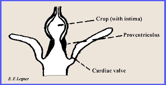
This is also the main site of secretion. Morphology (Digestive Tract). -- The digestive tract
is divided into the Stomodaeum,
Mesenteron
and Protodaeum. A simple diagram of the tract may be seen
in Fig. ent24.
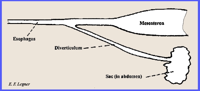
The Stomodaeum (or foregut) contains the esophagus, which is an
undifferentiated tube for simple conduction of food. The crop follows this, which is a storage organ. It may serve for partial digestion,
especially sugar and starch, as fluids from the mesenteron pass forward into
the crop. In some insects, e.g.,
mosquito, the crop may appear in the form of a diverticulum.
The Salivary
Gland empties from the anterior portion of the insect near the
mouth. It is a very active structure,
and the saliva is used in digestion and as an anticoagulant in some groups
(e.g., aphids). It also may be
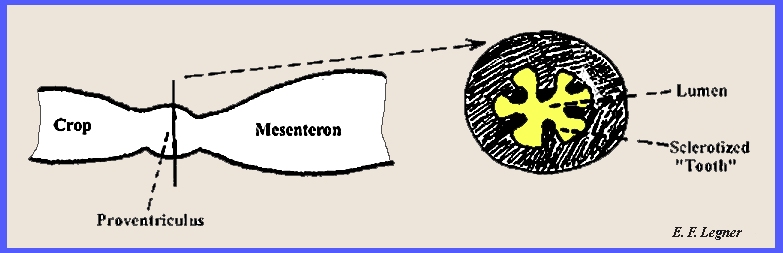
modified as in the silk-secreting organ of Lepidoptera. The proventriculus follows the crop and may be a secondary masticating organ,
as in the cockroach. It actually
serves as a food strainer.
The cardiac valve keeps the food moving in a posterior direction.
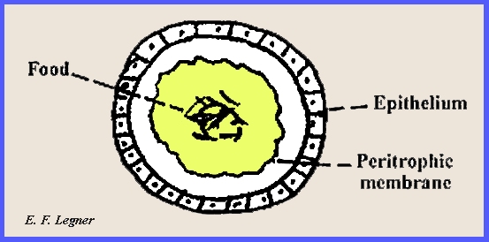
The Mesenteron (or midgut) is the site where nearly
all digestion occurs, which is primarily fat and protein. Most nutrients are absorbed here. The cells are extremely active and there
is a continual replacement of cells in the lining. The gastric caecae are expansions of the
midgut (Fig. ent24). The peritrophic membrane is secreted by epithelial
cells, which are either isolated groups of cells or by all the cells of the
midgut. It is permeable to liquids
and also may serve as a protective device for tender tissue of the stomach.
The Proctodaeum (or hindgut) is the
principal site for water absorption.
It contains a pyloric valve, and the Malpighian tubules that serve for excretion
enter the hind gut at the junction of this valve. The rectal sac at the end of the
digestive tract is very important in water absorption for most insects. However, some groups such as clothes moths
utilize metabolic water. Excretion. -- Hypodermal cells of the integument absorb waste products and
secrete them into the cuticle, which is shed at the molt. However, the Malpighian tubules serve as
the principal excretory structure. Fat Body. -- Many insects cannot feed in the adult stage; hence their energy
as adults comes from the fat body, which is built up during immaturity. ------------------------------------------- A simple diaphragm shows the
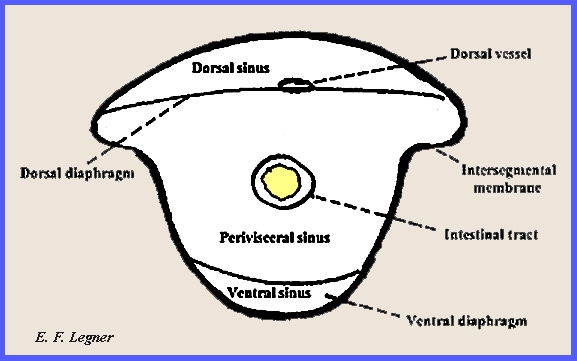
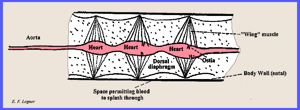
principal parts of the Insect circulatory system (ent29):
Diaphragms.
-- These consist of sheets of muscle that are connected together by
membranes. The dorsal diaphragm is
usually present, but the ventral diaphragm is variable in occurrence. Dorsal
Vessel. -- There are several
hearts, which are provided with circular muscles for contracting and
expanding. They serve as pulsating
organs. The blood is propelled
forward usually, but sometimes it may go backwards. Auxiliary pumping membranes sometimes occur at the bases of the
extremities, and they pump blood to those areas (e.g., legs, antennae, etc.).
Blood.
-- Insect blood is a viscous liquid that consists of about 75 percent
water. Pigmentation varies from clear
to red, brown or yellow. The oxygen
concentration is never very high in insect blood, so the blood does not serve
as an efficient oxygen carrier.
Amoeboid cells (phagocytes) occur, which engulf foreign materials and assist
in clotting. Clotting is mainly a coagulation
of phagocytes and there is no fibrinin.
In most insects this clotting mechanism is very efficient. ------------------------------------------- As noted the insect blood does not
serve as a principal vehicle for the transmission of Oxygen. There are other ways that this is
accomplished. Diffusion Through The Integument.
-- For this to occur the integument must be thin and wet at all times. Gill filaments or plates may be involved
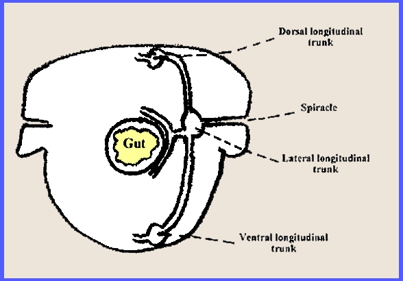
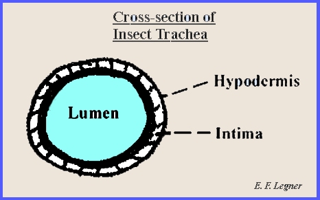
and parasitic forms obtain their oxygen directly through the integument. Tracheal
System. -- This is a complicated
system of tubes, which extends all through the insect body. The individual tubes are called tracheae. Entrance with the external environment is
via the spiracles.
Internections have occurred between the
metameres to form a continuous system.
The tracheal system originates from invaginations of the body wall,
and at the molt the tracheae are also cast off.
Air sacs
may appear at any point on the trunks. Bracing of the tracheae is accomplished by
taenidia,
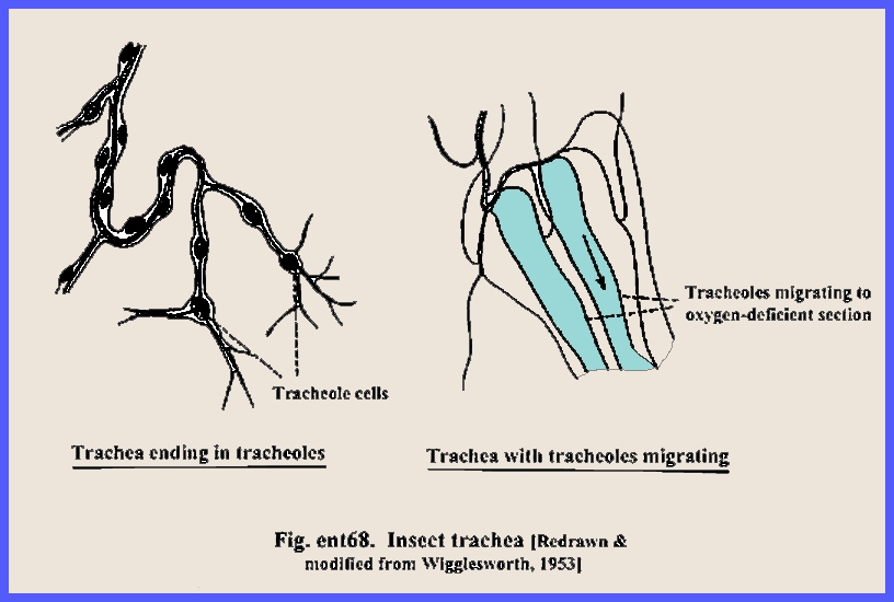
which occur in a spiral within. Tracheoles
are a series of
single-celled tubes that are located at the ends of the system. These cells lie on the surface of the
muscles and other body tissue. They
are not lined with intima and they are filled with a fluid, which serves as a
medium for gas exchange. Molting
includes a portion of the tracheoles.
A diagram of trachea and tracheoles in the system may be viewed in
Fig. ent68.
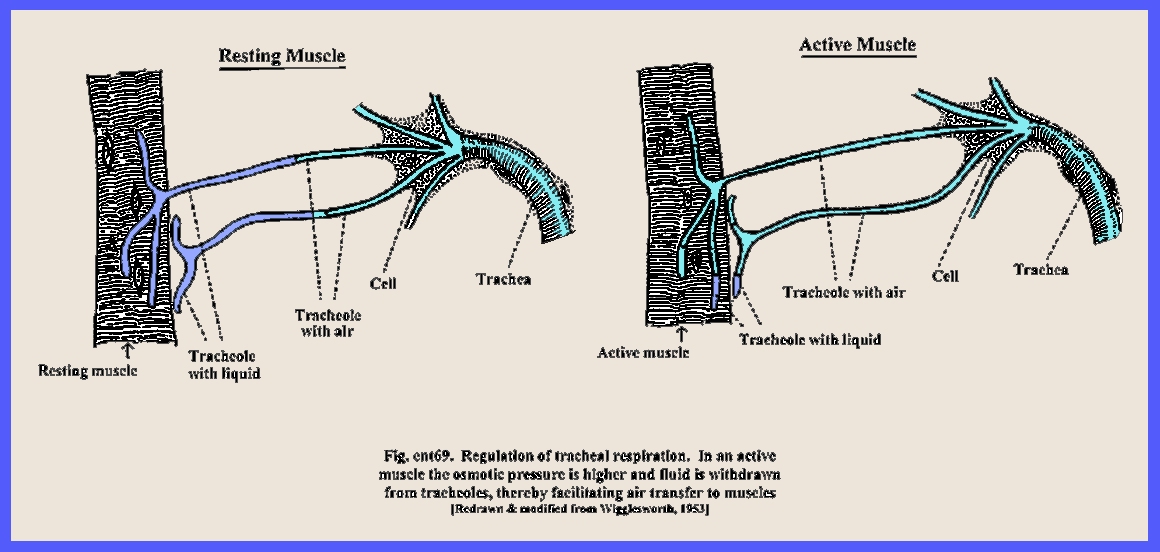
Body movements
augment the respiratory function. Figure ent69
shows how trachea transfer oxygen to the muscles:
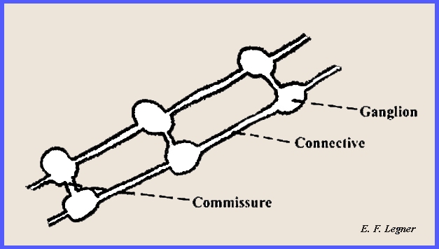
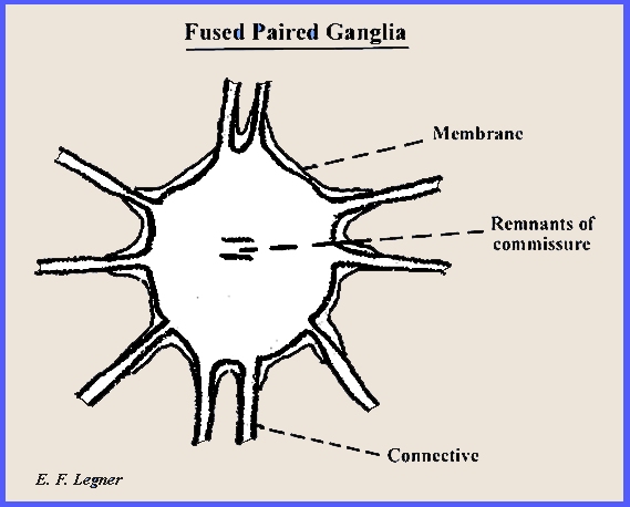
------------------------------------------- Ventral Nerve Cord.
-- Primitively there was a pair of ganglia for each segment. Modern insects have these ganglia fused
and the total number reduced.
There are never more than eight
ganglia in the abdomen, and then thee is a tendency for further reduction.
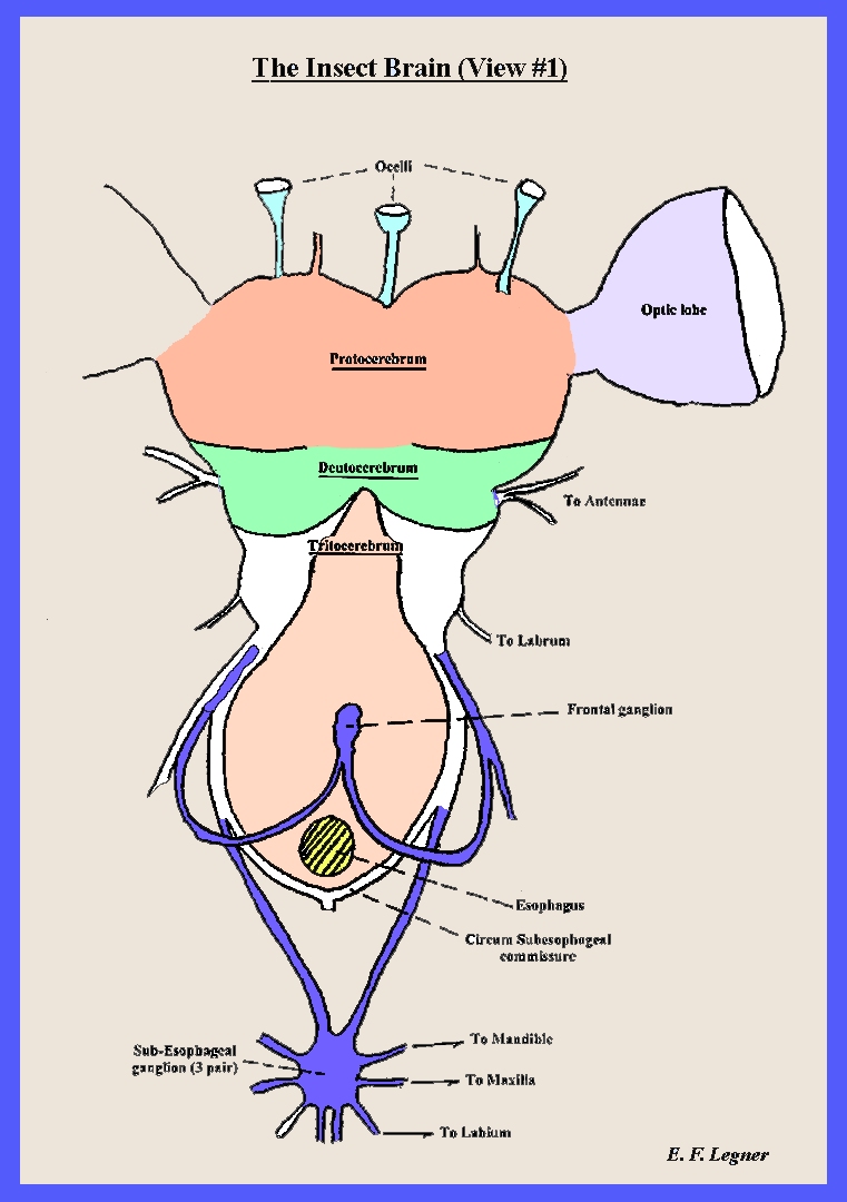
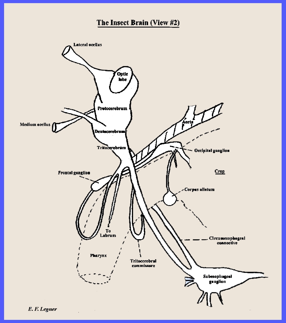
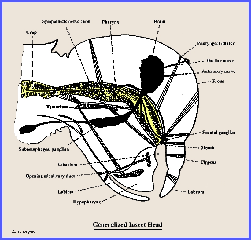
The
Insect Brain. -- This is primarily a
sensory area. Most body functions can
continue for a long time even when the head is removed (See Ent35 and Ent36 for
diagrams) The insect brain is divided into
three regions. (1) The Supraesophageal
Ganglion consists of a protocerebrum, a deutocerebrum
and a tritocerebrum;
(2) the Subesophageal
Ganglion is a fusion of three pairs of ganglia. Each pair enervates a portion of the
mouthparts; and (3) the Frontal Ganglion is connected to the tritocerebrum and forms
a part of the stomodael system.
Insect Ocelli.
-- The lens is simply a clear space in the integument. Light can enter at any angle, and hence
thee is no clear image produced. They
are sensitive to light intensity and may serve to detect motion.
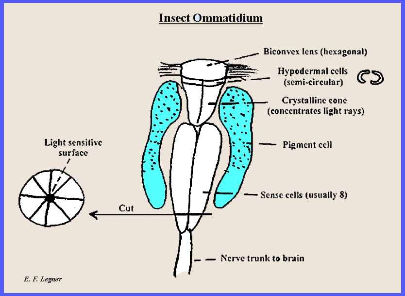
The
Compound Eye. -- Each lens is also a
clear space in the integument. A
crystalline cone lies beneath the cornea.
There are pigment cells that contain granules of pigment, which can
migrate up or down, depending on the light intensity. Light sensitive cells are located in an
octagon at the bottom of the cone. There are two opinions about what
the insect actually sees: (1) each
ommatidium sees a whole image, and (2) each ommatidium seas only a segment of
the image, thereby producing "mosaic vision."
Auditory
Organs. -- Some insects, such as
the grasshopper, possess a tympanum that allows hearing. But most insects hear through their
antennae. ------------------------------------------- All insects are unisexual, but when gynandromorphs
occur they are nonfunctional. Functioning
wise, many insects, such as worker bees, are asexual. In bees there is a suppression of the
sexes and only one female is singled out for reproduction. Secondary sexual characteristics are often present. This is illustrated by the frenulum in
moths, and the eyes of male flies are closer together than those of the
female. Diagrams of the male and female
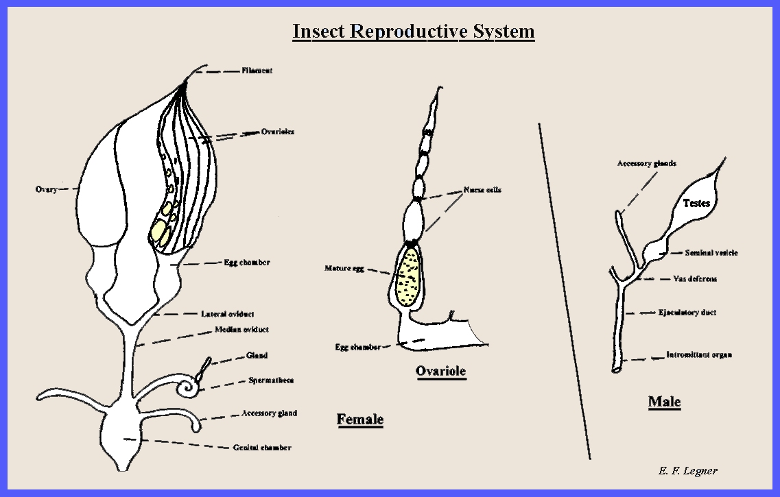
reproductive systems may be seen in Figure Ent40.
The Female Reproductive System.
-- There are two
ovaries, each consists of ovarioles and an ovary wall; a
filament attaches the ovary to the body wall, and nurse cells lie between the
ovules of the ovariole at varied sites. An Egg chamber receives a mature
egg from an ovariole. Additional structures are a lateral oviduct,
median oviduct,
genital
chamber, spermatheca with an accessory gland to nurse the spermatozoa
and accessory glands.
The latter serve for gluing the eggs to a surface and for pod
formation. The genital opening may occur on the
primitive 8th segment or it may have been shifted to the 9th segment. The Male Reproductive System.
-- The morphology of the male system consists of two testes, a seminal vesicle,
a vas deferens,
accessory
glands that furnish a matrix for passage of the sperm, an ejaculatory
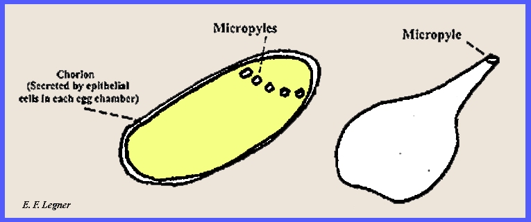
organ and an intromittant organ. Fertilization.
-- Spermatozoa enter the egg by way of minute pores in the chorion called micropyles. A tube may then extend down to the
interior of the egg.
------------------------------------------- The muscular system is quite complex,
consisting of from hundreds to several thousand individual muscles. They are all striated muscle cells, even
those that surround the alimentary canal and the heart. Insect muscles are able to
contract very rapidly and because of this they are very efficient, as such
action requires and adequate oxygen supply.
This is supplied by the tracheal system. The antennae of insects have
muscles only in the proximal end, with a few exceptions among the beetles
(Coleoptera). There is no internal
musculature in the wings, mandibles and some other structures. However, the legs and thorax have a
well-developed musculature. Thorax Musculature.
-- The various structures associated with muscles that occur in the thorax
are discussed as follows: (1) Phragma
are apodemes that extend
down from the dorsal surface of the integument. They are used for muscle attachment. (2) Dorsal
Longitudinal Muscles
arch the tergum and make
down strokes of the wing. In the
prothorax they extend to the head.
(3) Dorsal
Ventral Muscles
depress the tergum and are
for producing upstrokes of the wing.
(4) Furcae
are ventral apodemes for
muscle attachment. (5) Lateral or oblique muscles are
present. (6) Ventral
longitudinal muscles
are also present. (7)
Additional smaller muscles occur whose function is
debatable. The
Insect Head. -- Figure ent65 shows a
generalized diagram of the insect head.
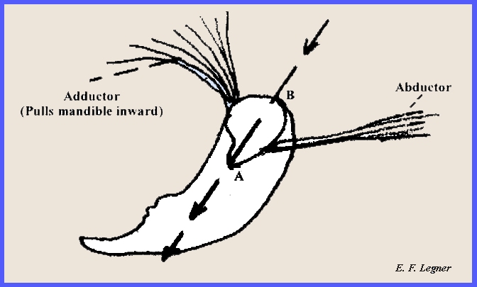
The
Mandible. -- The two mandibles are
attached at two points on the head (See Figure Ent41: A
& B). There are no muscles within the mandible
itself. The adductor muscle in the head
pulls the mandible inward while the abductor muscle extends the
mandible.
The
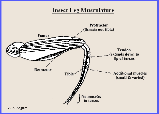
Insect Leg. -- Muscles extend from
the coxa down through the tibia, but there are none in the tarsus. The femur has a protractor muscle, which thrusts out the tibia and a retractor muscle
that pulls it back. The tibia
has some small and varied
muscles and a tendon
that extends down to the tip of the tarsus. (See Figure Ent42)
The Insect Abdomen.
-- Figure ent66 gives a diagram of the dorsal
part of the abdomen, showing various parts and muscles.
------------------------------------------- ============= |
