File:
<acanthocephala.htm> <Index to Invertebrates> <Bibliography> <Glossary> Site Description
<Navigate to Home>
|
Invertebrate
Zoology Kingdom: Animalia, Phylum: Acanthocephala (Contact)
CLICK on underlined file names and included illustrations to
enlarge: The Phylum Acanthocephala derives its name from
"spiny-headed." All species
are parasites in the digestive tract of vertebrates with intermediate hosts
that are generally arthropods. The
anterior end of the animal has a probosis with spiny hooks. A representative genus is Macracanthorhynchus
with characteristics noted as follows: General
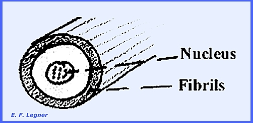
Body Features.-- A retractable
proboscis is present. There is a
genital pore at the posterior end, which in the female is very simple and in
the male is an inflatable fan-like affair or bursa
that is used for copulation. The
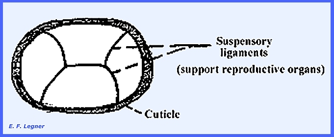
female is larger than the male. Body Wall.-- A heavy
cuticle is present. Underneath the
cuticle and secreting it lies the epidermis.
All cell membranes are lost between the cells, a condition that is
known as a syncytium. In the syncytium nuclei are scattered
through an undivided mass of cytoplasm.
Both circular and longitudinal muscles are well developed and lie
under the epidermis. The muscle cells
consist of tapering hollow cylinders .
Pseudocoelom.--
This structure differs from a true coelom in not being derived entirely from
the mesoderm. In a true coelom the
body cavity is lined on all sides by mesoderm. However, it serves the same purpose as a true coelom. It takes up practically all of the
interior of the animal. Inside the
pseudocoelom there is a fluid, which maintains turgor. Food
& Digestion.-- There is no mouth
or any sign of a digestive tract, and thus the animals are similar to the
Cestoda. Predigested food is absorbed
through the body wall. Respiration.--
The animals respire by diffusion and it is anaerobic for considerable
periods. Excretion.--
Flame cells, which are not scattered, accomplishes excretion. They are gathered into two large tufts
projecting out into the pseudocoelom and emptying out through the
reproductive tract (= urogenital system). Nervous System.--
A simple nervous system is present consisting of a single ganglion, which
lies on the outer wall of the proboscis sheath. Various nerves run out to different parts of the body
particularly to the muscles. This is
then much simplier than that of the Turbellaria. Reproduction.-- Extending
the whole length of the body and dividing up the pseudocoelom in sections are
suspensory ligaments. The male has two testes attached to a
suspensory ligament, and the vas eferens and vas deferens are present. There are a series of 6-8 cement
glands
on the sides of the vas
deferens, which lead back to the copulatory bursa that
serves an adhesive funtion during copulation.
The female has a series of ovaries
attached to the suspensory ligaments.
Floating ovaries
are present, which are
little patches of ovary tissue that break off of the main ovary and bear the
maturing ova. The eggs when mature
detach from the ovaries and they too float in the fluid of the
pseudocoelom. Eggs are fertilized in
the pseudocoelom by the sperm that enter the cavity. Eggs with developing embryos bear 2-3
heavy shell layers. At the opening of
the female genital tract is a remarkable structure, the uterine
bell, which is a selective
apparatus.
Fluid of the pseudocoelom is
sucked into the mouth of the apparatus.
A selection of mature eggs is made at the base and immature eggs are
passed back into the pseudocoelom.
The mature eggs are laid outside following selection that is made on
the basis of size and shape of the egg. Lemnisci.--
These are glandular structures, which occur at the anterior end of the
animal. They are part of the lacunar
system that runs all through
the epidermis. They may serve as
reservoirs for fluid, which moves around the lacunar system. Cell
Constancy.-- There is a definite
number of cells per each organ, thus the number of epidermal, muscular, nerve
cells, etc. is constant per species.
This results in a cessation of cell division in the late embryonic
period of development, except for the ovaries and testes. All further increase in size is due to the
enlargement of existing cells.
Various parts of the body consist of definite numbers of cells (except
the gonads). Along with this there is
a tremendous increase in the size of the nuclei. A syncytium frequently results. Life
History. -- Macracanthorhynchus is
a parasite of pigs. The eggs pass out
with the host's faeces. June beeetle
larvae consume the eggs and the embryo leaves the egg and bores through to
the body cavity of the beetle. The
hog must devour the beetle to complete the life cycle. Different species of parasite have
different intermediate hosts of aquatic insects and crustaceans, etc. Importance.--
There is relatively little economic importance although infection by Macracanthorhynchus may lower
the general health of pigs if present in large numbers. ------------------------------------ Please see
following plates for Example Structures of the Acanthocephala: Plate 29 = Phylum: Acanthocephala: Macracanthorhynchus
sp. male Plate 30 = Phylum: Acanthocephala: Macracanthorhynchus
sp. -- Cross-sections Plate 31 = Phylum: Acanthocephala, Macracanthorhynchus
sp. -- Dissected Female specimen ============== |