`File: <fly-par.htm> General Index Terminology [Navigate to
MAIN MENU ]
|
KEY TO ADULTS OF PRINCIPAL PARASITOIDS OF SYNANTHROPIC DIPTERA BREEDING IN DECOMPOSING ORGANIC WASTES 1 E. F. Legner University of California Riverside, California ----Please CLICK
on desired underlined categories [ To search for
Subject Matter,
depress Ctrl/F ]: See
MORPHOLOGY to learn about insect
structure Details
on families may be found in <taxnames.htm> Records of the activity of
parasitoids from synanthropic Diptera developing in decomposing organic
wastes were repetitious enough by 1976 from collections sites throughout the world
to suggest that most major species were known (Girault 1910, Girault &
Sanders 1910, Howard 1911, Hewitt 1914, Graham-Smith 1916, 1919; Bridwell
1919, Froggatt 1921, Johnston & Bancroft 1920, Johnston & Tiegs 1921,
Séguy 1923, Vanderburg 1929, 1930, 1931; Newman & Andrewartha 1930,
Handschin 1932, Feng 1933, Lindquist 1936, Miller et al. 1936, Campbell
1938, Roy & Siddons 1939, Simmonds 1940, 1967, 1958; Thompson 1943, 1944;
Bromley 1945, Wolcott 1948, Muesebeck et
al. 1951, West 1951, West &
Peters 1973, Nikolskaya 1952, Steve 1959, Azab et al 1963, Boucek
1963, Peck 1951, 1963, 1974; Sytshevskaya 1963, 1964, Jenkins 1964 Peck et al.
1964, Yasumatsu & Watanabe 1964; Legner 1965a,b; 1966, 1967a,b,c; 1969b;
Legner et al. 1967, 1974a,b; 1976; Legner & Greathead 1969, Legner
& McCoy 1966, Legner & Olton 1968a,b; 1971, 1975; Legner &
Poorbaugh 1972; Houser & Wingo 1967b, Jones 1967a,b; 1971, Sanders 1967,
Sanders & Dobson 1966, Depner 1968, Beard 1969a, Combs & Hoelscher
1969, Mourier & ben Hannine 1969, Kogan & Legner 1970, Moore &
Legner 1971, Mourier 1971, Greenberg 1971, Anonymous 1972, Monty 1972, Thomas
& Wingo 1968, Thomas & Morgan 1972, Turner et al. 1968, Wylie
1973a, Ables & Shepard 1974a,b; 1976a; Keiding 1974, Mitchell et al.
1974, Ursu & Tudor 1975, Ursu et
al 1976). Thereafter, only a few additional parasitoids have been added
to the list which proportionally usually demonstrate infrequent
parasitization activity on their hosts (Geetha-Bai & Sankaran 1977, Watts
& Combs 1977, Wharton 1979, Murphy 1980, Rongsiryam et al. 1980, Rutz &
Axtell 1980b, Andriescu & Fabritius 1981, Butler & Escher 1981,
Butler et al. 1981, Dabbour et al. 1981, Thomas 1981, Figg et al.
1982, Murakami 1982, Fabritius 1983b, 1987a,b; Hulley 1983, Meyer & Petersen
1983, Meyer et al. 1990a, 1991; Geetha-Bai & Sankaran 1985, Rueda 1984,
Rueda & Axtell 1985a,b,c,; 1987, Rueda et al. 1990, Xue 1984b,
1986a, 1987a, 1988b; Xue & Zhang 1982, 1983, 1989; Zhang & Xue 1984,
Zhang et al. 1990, Smith & Rutz 1985, 1991b,c,d; Smith et al.
1989; Axtell & Rutz 1986, Keiding 1986, Mullens et al. 1986, Patterson
& Rutz 1986, Merchant et al. 1987, Skoda et al. 1987, Hoebeke
& Rutz 1988, Berti-Filho et al. 1989, Geden et al. 1989, Greene et al.
1989, Pinheiro et al. 1989, Rutz & Scoles 1989,
Huang 1990, Miller & Rutz 1990, Henderson & Rutz 1991, Blanchot 1992,
Klunker & Fabritius 1992). These parasitoids are believed to
play an important role in reducing the average density of their dipterous
hosts, frequently producing >90% mortality of the later host developmental
stages, with parasitoids attacking the pupal stage being predominant (Smit
1929, Simmonds 1948, Davis 1960, Saunders 1962, Graham & Harris 1966,
Keiding & ben Hannine 1966, Legner & Brydon 1966, Legner et al.
1966d, 1975, 1982; Legner & Greathead 1969, Legner 1970a,b; 1971, 1975,
1976a,b, 1977, 1978a,b; Legner 1983b, 1986a, 1986b,c; Legner & Bay
1970a,b; Legner & Olton 1971, Legner & Dietrick 1972, 1974; Legner
& Bowen 1973, Legner & Badgley 1982, Legner & Warkentin 1985,
Waterhouse & Wilson 1968, Mourier & ben Hannine 1969, Mourier 1972,
Wright & Spates 1972, Nabrotsky 1974, Ables & Shepard 1975, Toyama
& Ikeda 1976, 1980; Lancaster 1979, Arditi 1980, Fabritius 1980c, 1981a;
Fabritius & Ursu 1990, 1981; Rutz & Axtell 1980a, Rutz &
Patterson 1990, Axtell 1981, 1986, 1990; Dietrick 1981, Drea & King 1981,
Harris 1981, Hogsette & Butler 1981, Morgan 1981a, Morgan & Patterson
1975b, 1986; Morgan et al. 1976c, 1988; Patterson et al.
1981, Greathead (1980, 1986), Petersen et
al. 1981, 1990; Petersen &
Meyer 1985, Petersen 1986a,b; Stage & Petersen 1981, Weidhaas 1981, 1986;
Weidhaas & Morgan 1977, Meyer & Petersen 1982, Meyer et al.
1990b, Morales-Pérez 1982,Murphy 1982, Ripa 1983, 1986a,b; 1990, Costello 1984,
Guzman 1984, Merchant et al. 1985, Fabritius 1986a,b; Fabritius
& Andriescu 1984, Guzman & Petersen 1986a,b; Meyer 1986, 1990; Meyer et al.
1990a, Rutz 1986, Rutz & Axtell 1979, Lazarus et al. 1989,Smith 1989,
Smith & Rutz 1991a, Xue 1990a,c; Zhang & Xue 1990, Zhang et al.
1990 Geden 1991, Geden & Rutz 1991a,b; Geden et al. 1992b, Klunker
& Fabritius 1991, Klunker & Kieson 1980, Wilhoit et al. 1991a,b). The degree of parasitization of Diptera
that breed in isolated field habitats such as animal dung, is low compared to
accumulated deposits, with few of the parasitic species involved being common
to those found in accumulated organic wastes (Olton & Legner 1973, Legner
et al. 1974a). The
characteristics of accumulated organic wastes cause an attraction for a
distinct dipterous as well as parasitic fauna, which may be related to a
higher humidity and reduced rate of decomposition. Therefore, accumulated wastes are also a primary producer of
Diptera of medical and veterinary importance such as the common housefly,
stable flies and several species of Fannia. It is well known that wild
parasitoid populations exhibit seasonal and geographical differences in
behavior and morphology. Therefore,
collections meant for importation should optimally include isolates from
diverse areas and different times of the year. Differences include aggressiveness, heat and cold tolerance,
and uniparentalism, gregarious versus solitary development, the number of
eggs deposited into a single host, larval cannibalism intensity and
parasitoid size. Detailed studies on Muscidifurax uniraptor, M. raptor and M. raptorellus demonstrate the
great amount of diversity that can be found within one genus. The following key and illustrations
are presented as a simplified means of identification of the principal
parasitoids of synanthropic Diptera breeding in decomposing organic wastes,
especially for those not familiar with hymenopteran terminology. Principal hosts include Musca domestica L., Stomoxys calcitrans (L.), Stomoxys nigra , Muscina stabulans (Fallen), Ophyra leucostoma (Wiedemann), Ophyra aenescens (Wiedemann), Fannia canicularis (L.), Fannia femoralis (Stein), Fannia scalaris (Fab.), and of
various species of Calliphora, Sarcophaga and Drosophila. In the preparation, considerable use was
made of works published by Borror et
al. (1981), Boucek (1963, 1965),
Boucek & Narendran (1981), Gauld & Bolton (1988), Gerling (1967),
Graham (1969), Kogan & Legner (1970, Legner et al. (1976), Nikolskaya
(1952), Peck et al. (1964), Riek (1970), Rueda &
Axtell (1985), Subba-Rao (1978), and Subba-Rao & Hayat (1985), and
Trjapitzin (1978). The parasitoids of
these flies continue to be exchanged around the world in biological control
efforts. (All Species see <flyparas.htm>) |
Please CLICK on following taxa for details.
|
(Ichneumonoidea) 2.
Alysia spp. 5.
Stilpnus
spp. (Chalcidoidea) |
9.
Dirhinus himalayanus Westwood 10. Tachinaephagus
javensis Subba-Rao 11. Tachinaephagus stomoxicida
S.-R. 12. Tachinaephagus
zealandicus Ashmead 13. Muscidifurax raptor
Girault & Sanders 14. Muscidifurax raptorellus
K. & L. 15. Muscidifurax raptoroides
K. & L. 16. Muscidifurax uniraptor
K. & L. 17. Muscidifurax zaraptor
K. & L. 18. Nasonia
vitripennis (Walker) 19. Pachycrepoideus
vindemiae Rondani 20. Spalangia
cameroni Perkins 21. Spalangia
drosophilae Ashmead |
24.
Spalangia longepetiolata Boucek 27.
Spalangia nigroaenea Curtis 28.
Spalangia rugulosa Förster 30.
Urolepis rufipes (Ashmead) (Cynipoidea) Cynipidae
(rare) 31. Figites spp. 32. Hexacola
(= Trybliographa) spp. (Proctotrupoidea) 33. Trichopria
spp. |
KEY TO
ADULTS OF PRINCIPAL PARASITOIDS OF SYNANTHROPIC
DIPTERA
BREEDING IN DECOMPOSING ORGANIC WASTES [This key is in a form commonly used in North
America. If the statement is true,
proceed to the designated
couplet, whereas if it is false, go to the "b" portion of the
couplet. Numbers in
parentheses refer to previous couplet or couplets read]. ----Please
CLICK on desired underlined categories to view pictures and to navigate in
the key [A will display all pictures for both
pairs of a couplet] ---------------------------------------------------------------------------------------------------------------------------- |
|
(Chalcidoidea)
- Chalcididae:
(Chalcidoidea) - Encyrtidae:
(Chalcidoidea) - Pteromalidae:
Muscidifurax
spp.
Spalangia spp.
(Chalcidoidea) - Encyrtidae:
(Chalcidoidea)
- Chalcididae (Dirhininae):
(Cynipoidea):
(Ichneumonoidea):
(Ichneumonoidea) - Ichneumonidae
(Proctotrupoidea)
- Diapriidae:
Staphylinidae:
[End of Key] |
|
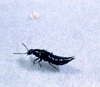
================================================================================== DESCRIPTIONS OF SPECIES Parasitic Staphylinidae (Coleoptera) The
front wings are without veins, hard or leathery, short, exposing much of
abdomen (Fig. 49a-A).
The first visible abdominal sternite is not interrupted by hind coxal
cavities. Outer lobe or galea of maxillae
not a segmented process. Maxillary
palpi are much shorter than antennae.
Hind tarsi with at least as many segments as front and middle tarsi. Two important genera are Aleochara and Anotylus. Key references are Thompson (1944),
Peschke & Fuldner (1977), Seevers (1978), Borror et al. (1981), Peschke et al.
(1987a), and Omar et al. (1991a) 1. Aleochara spp.
(Coleoptera: Staphylinidae: Aleocharinae) (PHOTO) These parasitoids are characterized by the forewings
being developed into leathery shields (elytra) under which the hind wings
(the organs of flight) are folded when at rest. The elytra are short, leaving much of the abdomen exposed (Fig. 50a-A & B). The abdomen is highly
flexible as in Aleochara
taeniata
Erichson (Fig. 50a-C). The genus Aleochara differs from other staphylinids in that the antennae are
inserted on the face between the anterior margins of the eyes, the tarsi have
5 segments, the maxillary palpi have 5 segments and the labial palpi have 4
segments. The two terminal segments
of the palpus are much narrower than the preceding with the last segment
minute. All species of this genus in
which life histories are known are solitary ectophagous parasitoids on the
pupae of muscoid flies within the puparium (Kemner 1926, Legner et al.
1976, Lesne & Mercier 1922, Jones (1967), Moore & Legner 1971, 1973,
1974a,b; 1975, White & Legner 1966).
Additional key references are Coquillett (1891), Wadsworth (1915), Scott
(1920), Kemner (1926), Soring (1927), Zorin (1927), Scheerpeltz (1933, 1934),
Burks (1952), Read (1962), Fuldner (1963, 1964, 1968, 1971, 1973), Drea
(1966), Horion (1967), Wingo et al. (1967), Allee (1969), Adashkevich
(1970), Adaskevich & Perekrest (1973, 1974), Hünten (1971), Riegel
(1971), Schneider (1971), Heller (1974, 1976), Heller & Treece (1976),
Lohse (1974), Pfenning (1975), Schulz (1975), Watts & Combs (1975),
Peschke & Fuldner (1977), Peschke & Metzler (1987), Ienistea & Fabritius (1978, 1982),
Kirknel (1978), Peschke (1978a,b,c; 1983, 1985a,b; 1986, 1987), Peschke et al.
(1987b), Ursu & Sperantia (1978), Tawfik et al. (1980),
Hertveldt et al. (1984a,b), Klimaszewski (1984), Klimaszewski & Blume
(1986), Klimaszewski & Cervenka (1986), Hunter et al. (1985),
Samsoe-Peteresen (1985, 1987), Whistlecraft et al. (1985), Gordon
& Cornect (1986), Scott & Rutz (1988), and Wright & Müller (1989). Family Braconidae (Hymenoptera: Ichneumonoidea) The fore wing costal cell is absent. There is either one recurrent vein (Fig. 45b-A)
or none. Ventral abdominal segments
are soft and membranous, with a median fold.
Abdomen is not much elongated and the propodeum is not prolonged
beyond hind coxae. These parasitoids
are less than 12 mm long (Fig. 45b-B) Two important genera are Alysia and Phaenocarpa. Key references are Marsh & Altson
(1920), Lima (1960, 1962), Tobias (1962, 1963), Riek (1970), Prince (1976),
Richards (1977), Borror et al. (1981), Zhao (1984), Wharton
(1976, 1986, 1987), Krombein et al. (1979), Gauld & Bolton (1988),
and Goulet & Huber (1993). 2. Alysia spp.
(Hymenoptera: Braconidae) The parasitoids of this genus are common in areas
of moderate to high rainfall (Myers 1927, 1929; Laing 1937, Griffiths 1964,
1966; Fischer 1970, 1971; Wharton 1986).
A common species in Europe, A.
manducator
Panzer, was successfully established in Australia and New Zealand (Miller
1927, Newman 1928, Morgan 1929, Holdaway & Evans 1930, Holdaway &
Smith 1932.) The nearctic Alysia ridibunda
Say has been found active on Calliphoridae (Lindquist 1932, 1940; Marsh
1968). Species of Alysia may be distinguished from those of Aphaereta by their larger size (ca. 2X as long), their shining black body
color and the uneven thickness of antennal funicular segments. They are solitary endophagous larval
parasitoids of muscoid flies. Additional key references are Altson (1920),
Caudri (1941), Likovský (1965, 1973), Burgess & Wingo (1968), Vinogradova
& Zinovjeva (1972), Zinovjeva (1976, 1978, 1981, 1985, 1987, 1988),
Chernoguz (1984, 1986), Chernoguz & Reznik (1987), Chernoz & Vaghina
(1987), and Chernoguz et al. (1987). 3. Aphaereta spp.
(Hymenoptera: Braconidae) These parasitoids are found primarily in
climates with substantial rainfall. Aphaereta pallipes
(Say) is most frequently encountered in the Holarctic (McComb 1958, Salkeld
1959, House & Barlow 1961, Griffiths 1964, 1966; Lange 1964, Lange &
Bronskill 1964, Houser 1966, Houser & Wingo 1967a, Marsh 1969, Fischer
1970, 1971; Garry & Wingo 1971, Figg et al 1983a,b, Whistlecraft et al.
1984, Rueda & Axtell 1985b). Aphaereta aotea Hughes &
Woolcock is found in Australia (Hughes & Woolcock 1976, 1978, Hughes et
al 1974). Species of Aphaereta may be readily distinguished from those of Alysia by their smaller size (ca. 1/2 as long), their reddish-brown
body color and uniform thickness of funicular antennal segments. They are solitary endophagous larval parasitoids
of muscoid flies. Key references are
Fischer (1966), Zinovjeva (1974), and Gherasin & Lacatusu (1977). Family Ichneumonidae (Hymenoptera: Ichneumonoidea) Forewing venation is complete, not reduced,
with at least one complete cell present.
Stigma and costal cell are absent.
There are 2 recurrent veins usually present (Fig. 45a-A & B). Ventral abdominal
segments are soft and membranous, with a median fold. Two important genera are Phygadeuon and Stilpnus. Key references are Thompson (1944), Salt
(1952), Townes & Townes (1966, 1973), Riek (1970) Richards (1977),
Krombein et al. (1979), Borror et al. (1981), Subba-Rao & Hayat
(1985), Pisicà & Fabritius (1986), Blanchot (1988, 1991a,b), Gauld &
Bolton (1988), Rollard (1988) and Goulet & Huber (1993). 4. Phygadeuon spp.
(Hymenoptera: Ichneumonidae) This genus and Stilpnus can be distinguished from other parasitoids noted herein by
their complete wing venation. Both the
fore wings and hind wings have venation closed to form several cells (Fig. 46a-A). This genus is unique here in having
antennae of 22 segments with the first two segments short and the third
segment longer than the first two combined.
The antennae are inserted in the middle of the face between the
eyes. The species are solitary
internal larval parasitoids most often found in humid higher Holarctic
latitudes (Legner 1966, Legner & Olton 1968, Legner et al. 1976). Additional key references are Monteith
(1956), Horstmann (1967, 1972, 1975, 1986), Müller (1971), Frilli (1973),
Plattner (1975, 1979), Plattner & Naton (1975), and Naton (1983). 5. Stilpnus spp.
(Hymenoptera: Ichneumonidae) The wing venation is complete as in Phygadeuon, there being complete cells in both the fore wings and hind
wings. Antennae have 16 segments with
the first two segments short, the first being shorter than the next two
together. The color is shining
metallic black. They are solitary
endophagous larval parasitoids, apparently restricted to the genus Fannia in accumulated organic wastes (Legner & Olton 1971, Legner et al.
1976, Loomis et al. 1968). They vary greatly in size with males being about 1/2 that of
females. A common species is S. anthomyiidiperda
(Viereck). Family Chalcididae (Hymenoptera: Chalcidoidea) Hind femora are greatly enlarged, ventrally
toothed (either a few large or many small teeth) (Fig. 18a-A). The prepectus is reduced
or fused, not triangular (Fig. 18a-B). The frons is projected into 2 horns
(around antennae) when viewed dorsally.
They are solitary parasitoids which attack mature host larvae or young
pupae. Their general appearance is
shown in Fig. 18a-E. Two important genera are Brachymeria and Dirhinus. Key references are Ashmead (1899),
Schmiederknecht (1907, 1909), Froggatt (1916), Thompson (1944), Schmitz
(1946), Boucek (1956), Nikolskaya (1960), Riek (1970), Richards (1977), Krombein et al.
(1979), Subba-Rao & Hayat (1985), Fabritius & Andriescu (1987), Xue
(1988a), Xue et al. (1987c), Gauld & Bolton
(1988), and Goulet & Huber (1993). Genus Dirhinus
Dalman, 1818 (Hymenoptera: Chalcididae) (Dirrhinus Dalman, 1923; Eniaca Kirby, 1883; Dirrhinoidea Girault, 1912; Pareniaca Crawford, 1913; Eniacella Girault, 1913; Eniacomorpha Girault, 1915; Dirhinoides Masi, 1947). Parasitoids in this genus possess a pair of
horns on the head which in some species bear a tooth (Figs 42a-B
& D). They have an elongated
body which is somwhat depressed dorsally (Fig. 42a-E). The mandibles are long and narrow, almost
straight. The genae are very large
and punctured. They parasitize
various brachycerous Diptera, seeking out full-grown larvae or pupae in the
soil. The key reference is Boucek
& Narendran (1981). 6. Dirhinus anthracia Walker, 1846
(Hymenoptera: Chalcididae) (Dirrhinus
ruficornis Cameron, 1905; Eniacella
rufricornis
Girault, 1913; Eniacella bicornuticeps
Girault, 1915; Dirhinus sarcophagae
Froggatt, 1919; Dirhinus frequens
Masi, 1933; Dirhinus intermedius
Mani & Dubey, 1974). Female head densely punctate, in dorsal view with
eyes longer than temples, these converging, slightly convex (Fig. 42a-A). Antennae of female reddish with yellow or
white hairs. Each horn has a deep external
apical notch with subparallel ridged (= carinate) sides, the inner ridge
being laminate but almost regular (Fig. 42a-B
& C). Horn in middle is
usually distinctly broader than the gap between antennal sockets, in dorsal view. The ocellar area is somewhat elevated; in
lateral view the head is less than 0.6 as wide as long, in dorsal outline
from the horn edge to occiput it is strongly convex. However, the vertex appears as almost one
plane. Length of gena (from eye to submandibular
corner) is obviously greater than the short diameter of eye. Labrum bears scattered hairs. Antennal scape is thickened toward its
base. Pronotum bears small lateral
patches of thicker hairs, and the punctuation in center is dense. Abdominal petiole is transverse, area of 4
dorsal ridges about 2X as broad as long, anteriorly frequently margined. In male the horns dorsally in middle are
narrower than the gap. Size varies
with host size. The general
appearance is as in Fig. 42a-D
& E. There is a broad host range including
Muscidae, Sarcophagidae, Calliphoridae, Tephritidae and Tachinidae (Boucek
& Narendran (1981). Original
distribution India, Burma, Sri Lanka.
Additional key references are Walker (1846), Froggatt (1919, 1921),
Lever (1938), Masi (1947), Dresner (1954), Boucek (1956), and Mani et al.
(1974). 7. Dirhinus bakeri (Crawford, 1914) (Hymenoptera: Chalcididae) (Pareniaca
bakeri
Crawford, 1914; Pareniaca trichophthalma
Masi, 1927). This is a small species, 2.5-4.1 mm long, with
antennae usually black, but at times with pedicel and flagellar base in
female reddish. Facial edge of each
horn with a distinct additional tooth (viewed laterally) (Fig. 40b-A & B). Tip of horn reaches
much farther from eye than frontal tooth (Fig. 40b-A). Fore and mid femorae and tibiae usually
are black, and wings usually whitish.
Abdominal petiole is rather closely joined with abdomen. Gena of both sexes is longer than the
maximum diameter of eye (Fig. 40b-A). Hosts include Musca domestica
and species of Stratiomyiidae and Tachinidae (Boucek & Narendran
1981). Original distribution India,
Sri Lanka, Malaysia, Philippines, Japan.
Additional key references are Masi (1947), Habu (1960), Baltazar
(1966), and Geetha-Bai & Sankaran (1977). 8. Dirhinus banksi Rohwer, 1923 (Hymenoptera:
Chalcididae) Female body black including most of legs and
flagella. Scapes, pedicels, tarsi and
joints of legs are testaceous. Thorax
is much flattened dorsally, especially scutellum completely flat and with
broad smooth area separated by single row of punctuations from hind margin (Fig. 42b-A
& B). Lateral head at least 1.5X as long as wide, with facial
outline convex, and receding near horn tips.
In dorsal view head width is ca. 1.67X the minimum distance between
eyes, horns appearing wide and nearly with parallel sides in basal half (Fig. 42b-C). Flagellum plus pedicel 1.6-1.7 X as long
as head is wide in lateral view, and clava a bit shorter than 3 preceding
segments combined. Pronotum medially
without smooth longitudinal strip (Fig. 42b-B). Mesosternum with impunctate areas behind
the fore coxae frequently are well delimited from the posterior punctate part
and not extending quite 1/2 to mid coxae.
Petiole with area of 4 ridges slightly to moderately transverse. In male antennae are paler or darker
yellow, slightly less clavate than in female. Eyes are relatively small, in lateral view the height of eye is
only about 1.2X of the height of horn projecting above it. Length of female 2.5-3.7 mm., male 2.6-3.1
mm. The only known host is Lucilia sp. (Boucek & Narendran 1981). Original distribution India, Sri Lanka, Thailand, Cambodia,
Malaysia. Additional key references
are Rohwer (1923), Masi (1947), and Habu (1976). 9. Dirhinus himalayanus Westwood, 1836
(Hymenoptera: Chalcididae) (Dirrhinus
crythroceras
Cameron, 1906; Dirhinus luzonensis
Rohwer, 1923; Dirhinus luciliae
Rohwer, 1923; Dirhinus pachycerus
Masi, 1927; Dirhinus vlasovi
Nikolskaya, 1952; Dirhinoides mathuri
Mani & Dubey, 1972). Body is very black, with sparse coarse
punctuation on thorax; wings are clear without distinct hairs in female. The apex of each horn viewed dorsally is
almost rounded, without a notch (Fig. 41a-A
& B). Distal 1/2 of hind
tibia with another shallow groove outside tarsal sulcus, the groove being
outlined by some extra ridges. Clava
of female symmetrical, with a broad conical apex (Fig. 41a-C). Hind tibia have a conspicuous external ridge. Length of female 2.4-4.9 mm. (the longest
of all species noted here). Host
range is broad in Diptera in carcasses and excrement (Bouek & Narendran
1981). Original distribution Arabia,
India, Malaysia, Sumatra, Japan, Philippines & Hawaii. Additional key references are Cameron
(1906), Rohwer (1923), Ferrière (1935), Roy & Siddons (1939), Roy et al.
(1940, 1950), Stearn (1943), Nikolskaya (1952, 1960), Dresner (1954), Habu
(1960), Mani & Dubey (1972), and Sankaran (1977, 1985), Srinivasan & Panicker (1988), Xue
(1989), Geetha-Bai (1990), Geetha-Bai & Sankaran (1977, 1985), Family Encyrtidae (Hymenoptera: Chalcidoidea) Fore wing venation greatly reduced, with a
single vein along margin and very short spur. Fore wing with uncus well separated from postmarginal
vein. Marginal vein indistinct,
somewhat punctiform. All tarsi with 4
segments. Abdominal petiole at most
1-segmented. Eggs are
dumbbell-shaped. Endophagous,
gregarious larval parasitoids. The
principal genus is Tachinaephagus. Key references are Thompson (1944), Wilson
& Woolcock (1960), Legner & Bay (1965a), Riek (1970), Ho et al.
(1974), Richards (1977), Trjapitzin (1978), Krombein et al. (1979), Prinsloo
& Annecke (1979), Noyes (1980), Noyes & Hayat (1984), Borror et al.
(1981), Subba-Rao & Hayat (1985), Gauld & Bolton (1988) and Goulet
& Huber (1993). Genus Tachinaephagus Ashmead, 1904 (Hymenoptera: Encyrtidae) (Tachinaephagus
Girault, 1917; Australencyrtus Johnston & Tiegs, 1921; Australomalotylus Risbec, 1956) There are three species known to parasitize
synanthropic Diptera in decomposing organic wastes, although the whole genus
is exclusively parasitic on the larvae or pupae of many Diptera. Vertex of head is almost 1/3rd to 1/2 the
width of head, frons is broad. Eyes
are hairy with long dense setae.
Antennae inserted just below the lower eye level with toruli (=
sockets) widely separated, scape cylindrical, the funicle segments 1-3X
longer than broad (quadrate) (as in Fig. 38a-A). Mandibles have three sharp teeth. Scutellum long, shining but bears long setae
which arise from microscopic punctures.
Legs are hairy. Basal area of
fore wing is evenly setose (Fig. 39a-A). Abdomen is either slightly shorter or
longer than thorax and flat above in dead specimens. Digitus of male bears 3 teeth. The eggs are encyrtiform
(dumbbell-shaped). They are
endophagous gregarious larval parasitoids.
Size varies with host size and number of individuals developing on one
host. Key references are Tachikawa
(1963), Olton & Legner (1975) and Subba-Rao (1978). Other references are Ashmead (1904a,b),
Girault & Sanders (1909, 1910a), Girault (1917), Dodd (1921), Froggatt
(1921), Johnston & Tiegs (1921), Hardy (1924), Gourlay (1930a,b), Newman
& Andrewartha (1930), Ferrière (1933), Gahan (1938), Risbec (1956),
Ghesquière (1960), Olton (1971), and Subba-Rao (1972, 1976). 10. Tachinaephagus
javensis Subba-Rao, 1978
(Hymenoptera: Encyrtidae) Body is uniformly dark brown. Legs, coxae and antennal scape are
testaceous yellow, funicle and club brown; venation and discal cilia of
forewing brown. Head vertex is
broader than 1/2 the head width; ocelli are large in an equilateral triangle
(Fig. 38a-C), the posterior part separated from the ocular border by about
their own diameter. Antennal scape is
cylindrical, its pedicel only slightly longer than the 1st funicle segment,
2nd and 3rd segments shorter than 1st, 4-6 about equal but shorter than 3rd;
club apically rounded, the joints not well separated (Fig. 38a-A);
thorax is moderately convex; scutellum rugose; Fore wings with caudal cell
moderately wide, not parallel with submarginal vein (Fig. 38a-B);
marginal fringe short; abdomen longer than thorax, tergites shining and
smooth. Males have not been
found. Hosts include Haematobia and species of Musca (Subba-Rao
1978). Original distribution
Indonesia. The key reference is
Subba-Rao (1978). 11. Tachinaephagus stomoxicida
Subba-Rao, 1978 (Hymenoptera: Encyrtidae) Body is almost black; head vertex with faint
metallic green reflections; coxae are dark brown, rest of legs brown with
somewhat darker tarsi; scape brown, funicle and club dark brown. Fore wings are slightly hairy (Fig. 39a-A);
head vertex more than 1/2 width of head; ocelli in an equilateral triangle,
the posterior pair separated from ocular borders by a little more than their
own diameter. Antennal socket is
broad, semicircular and shallow.
Malar sulcus impressed deeply only basally. Antennal scape cylindrical, slightly dilated above, funicle
segments well separated, the club segments deep and club apex angular (Fig. 39a-C). Thorax is only slightly convex with
scutellum almost flat; mesoscutum scaly, scutellum smooth and shining except
for tiny pits bearing long black setae.
Fore wings are long and narrow, costal cell very narrow and parallel
with submarginal vein (Fig. 39a-A & B). Discal ciliation is
coarse and dense. Abdomen is slightly
longer than thorax, almost quadrate, tergites smooth and shining. Males resemble female except for their
antennae. The only known host is Stomoxys nigra (Subba-Rao
1978). Original distribution in
Uganda but established in Mauritius.
Key references are Subba-Rao (1978) and Greathead (1986). 12. Tachinaephagus
zealandicus Ashmead, 1904
(Hymenoptera: Encyrtidae) (Tachinaephagus
australiensis
Girault, 1917; Stenosterys
fulvoventralis
Dodd, 1921; Australencyrtus
giraulti
Johnston & Tiegs, 1921; Australomalotylus
rageaui
Risbec, 1956). The wing venation is greatly reduced with a
single vein along the margin and a very short spur, the stigmal vein, near
its center (Fig. 39b-B). Costal cell of forewing
wide, its border not parallel with submarginal vein (Fig. 39b-B & C). The antennae are
located in the middle of the face between the eyes. They are of less than 14 segments with the first segment
elongated, longer than the next two combined. As in Muscidifurax,
the pronotal disc is finely reticulate and almost imperceptibly
punctured. The color is shining black
with the underside of the thorax and the legs testaceous (Fig. 39b-D). Hosts include Calliphoridae, Fannia canicularis
(L.) and Musca domestica L. Original distribution
Australasia. Key references are
Tachikawa (1963), Olton (1971), Olton & Legner (1974, 1975) and Subba-Rao
(1978). Other references are Ashmead
(1904), Froggatt (1921), Johnston & Tiegs (1922), Hardy (1924), Gourlay
(1930a,b), Newman & Andrewartha (1930), Ferrière (1933), Gahan (1938,
Risbec (1956), and Legner & Olton (1968b). Family Pteromalidae (Hymenoptera: Chalcidoidea) Forewing marginal vein is less than twice as
long as stigmal (Fig. 19b-A). Antennae have 6 or
fewer funicle segments. Hind tarsus
with at least 5 segments. Abdominal
petiole often conspicuous and with dorsal carinae. They are ectophagous, pupal parasitoids inside the host
puparium. Important genera are Muscidifurax, Nasonia,
Pachycrepoideus,
Spalangia,
Sphegigaster,
and Urolepis. Key references are Latrielle (1805),
Dalman (1820), Walker (1836, 1839), Curtis (1839), Förster (1841, 1956),
Ashmead (1896b), Dalla-Torre (1898), Perkins (1910), Waterston (1915),
Fortsetzung (1916), Girault (1916, 1921), Parker (1924), Parker &
Thompson (1928), Ceballos (1941), Thompson (1944), Delucchi (1955), Boucek
(1963), Peck (1963), Baltazar (1966) DeSantis (1967, 1979, 1980), Graham (1969),
Riek (1970), Abraham (1975, 1978a), Wylie (1976b), Richards (1977), Burks
(1979), Gordh et al. (1979), Krombein et al.
(1979), Barlin & Vinson (1981), Borror et al. (1981),
Yoshimoto (1984), Subba-Rao & Hayat (1985), Gauld & Bolton (1988),
Hoebeke & Rutz (1988), Delvare & Aberlenc (1989), Grissell &
Schauff (1990), Goulet & Huber (1993). Genus Muscidifurax Girault & Sanders, 1910 (Hymenoptera:
Pteromalidae) Wing venation is incomplete and the marginal
vein is about twice as long as the stigmal vein (Fig. 23a-A). Antennal insertions are in the middle of
the face between the eyes. The first
antennal segment is longer than the next two combined, and there are less
than 14 antennal segments. Females
have one ring segment and 7 funicular segments (Fig. 23a-B),
males have 2 and 6, respectively. The
disc of the pronotum and the head are finely reticulate, without coarse
punctures. The several species are
very similar in appearance but have good behavioral characters distinguishing
them (Legner 1969a,b; Legner et al. 1976, Kogan & Legner 1970),
and they are electrophoretically distinct (Kawooya 1983). Females are black; males black with
translucent testaceous spots on the first, second and third ventral abdominal
segments. The eggs are
hymenopteriform, covered with small tubercles that distinguishes them from
those of Spalangia
(Gerling 1967) and with size differences for some species. The species may be either solitary or
gregarious. The average mass of
solitary species of this genus is relatively fixed, as host size does not
appreciably affect them (Legner 1969a).
They are ectophagous pupal parasitoids. The key reference is Kogan & Legner (1970). Van den Assem & Povel (1973) discussed
courtship behavior patterns that are specific. Markwick (1974) and Markwick et al. 1989 gave biological
characteristics that distinguish M.
raptor and M. zaraptor
and these species from Spalangia
endius. Other references referring to distribution,
identity, biology and genetics of species of this genus are Frison (1927),
Anonymous (1938), Nikolskaya (1952), Dresner (1954), Legner (1967b, 1969a,b;
1972, 1987a,b,c,d,e; 1988a, 1988b, 1988c,d; 1989a, 1990, 1991a, 1991b, 1993),
Legner & Dietrick (1974), Legner & Gerling (1967), Legner et al.
(1967), McCoy (1967), Wylie (1967, 1971a,b, 1972b), Berry & Speicher
(1972), Broadbent (1972), Kotschetova & Tjutjunkova (1973), Ables &
Shepard (1974), Kawooya (1983), van den Assem (1985), Propp (1986),
Mandeville & Mulles (1990b,c), Mandeville et al. (1990), Mann et al.
(1990b), Wilhoit et al. (1991a). 13. Muscidifurax
raptor Girault & Sanders,
1910 (Hymenoptera: Pteromalidae) (PHOTO) The fringe of setae (or their sockets) is well
developed on the posterio-apical margin of the fore wing (Fig. 23a-A). The stigma forms an abrupt enlargement at
the end of the stigmal vein, usually subquadrangular and distally acuminate where
a hair is often encountered (Fig. 27a-A). The uncus is directed distally. The frontal grooves are parallel (Fig. 27a-B). The median area of the propodeum of female
is closed behind (Fig 27a-C). The male digitus is
subtrapezoidal, broadest distally and usually with 3 apical processes (Fig. 27a-D). Hind legs with one tibial spur (Fig. 27a-E). Hind leg with tarsal claw bearing less
than 11 setae (Fig. 27a-F). Length of female is
2.33 mm, male 1.73 mm. The species is
biparental and solitary. An almost
cosmopolitan species which has not been collected in the Oriental region nor
most of Asia. Key references are
Girault & Sanders (1910a,c), McCoy (1963, 1967), Legner (1969a,b; 1976a),
Legner & Gerling (1967), Kogan & Legner (1970), Morgan &
Patterson (1975a), Fabritius (1978, 1981b,c,d; 1983a, 1984, 1986c) and
Kawooya (1983). Additional references are DeBach (1943), Sharma
(1967, 1971), Burton & Turner (1968), Shibles (1969), Wylie (1970),
Victorov & Azizov (1972), Markwick (1974), Tingle & Mitchell (1975),
Podoler & Mendel (1977), Fabritius (1979b, 1980a,b), Morgan et al.
(1979b), Capehart et al. (1981), Coch (1981), Klunker
(1981, 1982), Merritt et al. (1981), Rutz & Axtell (1981),
Shepard & Kissam (1981), Propp & Morgan (1983b, 1984a, 1985a), and
Geden et al. (1992a,c). 14. Muscidifurax
raptorellus Kogan & Legner,
1970 (Hymenoptera: Pteromalidae) The fringe of setae (or their sockets) is
absent from the posterio-apical margin of forewing, as in M. zaraptor
(Fig. 26b-A). The stigma may or may
not be elongated, sometimes being roundly clubbed. The uncus is directed towards of the apex of the wing. The pedicel of female antennae is not
slender proximally (Fig. 26b-B), and frontal grooves are usually convergent (Fig. 26b-C). The median area of the propodeum of female
is usually open behind with the lateral and median plicae not fused in the
middle (Fig. 26b-D). The male digitus is
subtrapezoidal, broader distally and usually with only 3 distal processes (Fig. 26b-E). Length of female is 2.11 mm, male 1.82 mm.
The Peru race averages female 2.33 mm, male 1.72 mm although they may assume
the smaller size of the Chilean race when deprived of host feeding. This South American species occurs as
several races demonstrating various degrees of gregarious development and
number of eggs deposited into a single host puparium. The Peruvian race is usually solitary in
development, but seasonal variations in larval cannibalism vary. The Chilean race has shown the greatest
degree of gregarious development (> 80%) and is quite thigmotactic. All known races are biparental. Key references are Kogan & Legner
(1970), Kawooya (1983), and Legner (1987b,e; 1988a,b,c; 1989a,b,c,d; 1990,
1991a,b; 1993). Superparasitism (= insertion of more
than one parasitoid egg per host) occurs in both the Peruvian and Chilean
race, and subsequent cannibalism by hatched larvae always follows. The Peruvian race deposits a lower number
of eggs per host than the Chilean race but cannibalism intensity is
higher. Therefore, it is difficult to
determine the exact number of eggs initially deposited by both species. The number of adult parasitoids that
survive is always less in the Peruvian race and usually averages about
one. On the contrary, more adult
survivors usually occur in the Chilean race, averaging about seven at a host
density of 20 per 24 hrs. A
standardization of host density, size, age and duration of exposure to
parasitization is essential in experiments as they influence the number of
eggs deposited and the rate of cannibalism. 15. Muscidifurax
raptoroides Kogan & Legner,
1970 (Hymenoptera: Pteromalidae) The fringe of setae (or their sockets) is well
developed on the posterio-apical margin of forewing. The stigma is formed as a gradual dilation
of the tip of the stigmal vein, and the uncus is directed distally (Fig. 28b-B). The frontal grooves are convergent. The male digitus is subrectangular and
usually with 4 apical processes (Fig. 28b-C). Spiracle of the female propodeum is not
remote from the lateral plica, i.e., the spiracular ridge is shorter than the
largest diameter of the spiracle (Fig. 28b-A). Length of female 2.31 mm, male 1.78
mm. This species is biparental and
solitary. It was originally known
from Costa Rica and southern Mexico.
Key references are Kogan & Legner (1970), and Legner et al.
(1976). 16. Muscidifurax
uniraptor Kogan & Legner,
1970 (Hymenoptera: Pteromalidae) The fringe of setae (or their sockets) is well
developed on the posterio-apical margin of forewing. The stigma is formed as a gradual dilation
of the tip of the stigmal vein, and the uncus is directed distally (Fig. 28a-B). The frontal grooves are convergent. When male are present, the digitus is
subrectangular and usually with 4 distal processes (Fig. 28a-C). The spiracle of the female propodeum is
remote from the lateral plica, i.e., the spiracular ridge is as long as the
longest diameter of the spiracle (Fig. 28a-A). Length of female 2.15 mm, male 1.68 mm,
the occasional male are the smallest in this genus. This species is uniparental and solitary in development. It was originally known only from Puerto
Rico where it occurs with the biparental M. raptor. Key references are Kogan & Legner
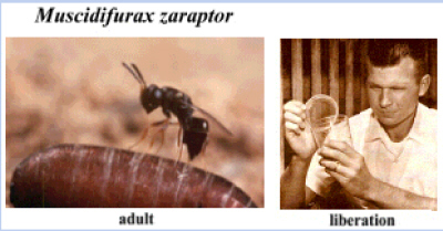
(1970), Legner (1985a,b; 1987a,d; 1988), and Kawooya (1983). 17. Muscidifurax
zaraptor Kogan & Legner,
1970 (Hymenoptera: Pteromalidae) The fringe of setae (or their sockets) is
absent from the posterio-apical margin of the forewing as in M. raptorellus. The stigma is small, elongated, suboval,
often acuminate at the internal angle where a hair is implanted (Fig. 26a-A). The uncus is usually directed toward the
anterior margin of the wing. The
antennal pedicel of female is obviously slender proximally (Fig. 26a-B),
and frontal grooves are usually parallel (Fig. 21a-C). The median area of the propodeum in female
is closed behind by the fusion of the lateral and median plicae (Fig. 26a-D). The male digitus is subrectangular with
4-5 apical processes (Fig. 26a-E). Hind leg with tarsal
claw bearing more than 11 setae (Fig. 26a-G). Length of female is 2.,84 mm, male 2.18
mm. This species is biparental,
solitary and occurs naturally in the western Nearctic. Key references are Kogan & Legner
(1970), Wylie (1971b, 1979), Legner 1977, 1979a,b,c) and Kawooya (1983). <PHOTO> Additional references are Coats (1976),
Petersen & Meyer (1983a,b), Petersen & Matthews (1984), Petersen
& Pawson (1988a,b; 1991), Petersen et
al. (1986, 1992), Pawson et al.
1987, Mandeville (1988), Mandeville & Mullens (1990a), and Mandeville et al.
(1988), Genus Nasonia Ashmead (Hymenoptera: Pteromalidae) 18. Nasonia vitripennis (Walker, 1836) (Hymenoptera: Pteromalidae) (Nasonia Ashmead, 1904; Mormoniella Ashmead, 1904; Pteromalus
vitripennis
Walker, 1836; Pteromalus muscarum
Hartig, 1838; Pteromalus abnormis
Boheman, 1856; Dicyclus pallinervosus
Walker, 1872; Stictonotus insuetus
Walker, 1872; Mormoniella brevicornis
Ashmead, 1904; Nasonia brevicornis
Ashmead, 1904; Platymesopus macellariae
Brèthes, 1913di). Head reticulate, wider than broad. Upper margin of antennal sockets is
slightly above level of the lower margin of compound eye. A distinct dorsocephalic ridge
exists. The malar sulcus is not as
pronounced as in Spalangia
endius (see Fig. 37a-D). Antennae are stout with two ring segments
and 6 funicular segments (Fig. 24a-B);
the pedicel is ca. 2 1/2X longer than first funicular segment. The pronotal collar bears an anterior
ridge and is strongly reticulate ventrolaterally. The mesosternum and scutellum is strongly reticulate, the
frenal groove is distinct (Fig. 24a-A). Propodeum has a median plica which lacks
lateral branches. The prosternum
bears a median groove. Fore wing with
marginal veins of uniform width and as wide as post marginal vein. Middle leg bears one tibial spur and dense
bristles over its entire length. The
spur is ca. 1/3rd as long as first tarsomere. The hind leg bears one tibial spur with a bristle-like
structure (Fig. 24a-C). Tergite 9 of abdomen
with a pair of slightly projecting sensory structures, each with 5 long
setae. Length of female 1.10-1.90 mm,
male 1.00-1.50 mm. Hosts are
primarily blowflies in animal carcasses, although occasionally parasitization
occurs in muscoid Diptera found in manure and accumulated organic
wastes. The species is
cosmopolitan. General appearance (Fig. 24a-E). Key references are Rueda & Axtell
(1985b). Other key references to this most extensively
studied (and undoubtedly the least beneficial) fly parasitoid include
Froggatt (1914a,b; 1918), Froggatt & McCarthy (1914), Robaud (1917),
Altson (1920), Miller (1922, 1972, Miller & Tsao 1974), Tiegs (1922),
Hardy (1924, 1925), Séguy (1929), W. Davies (1930), Dautert Willimzik (1931),
Cousín (1930, 1933), Evans (1933), Smirnov (1934), Smirnov & Kuzina
(1933), Smirnov & Vladimirow (1934), Vladimirova & Smirnov (1934),
Robaud & Colias-Belcour (1935), Fukuda (1939), Jacobi (1939), DeBach (1940),
DeBach & Smith (1941a,b; 1947), DeBach (1943), van der Merwe (1943),
Geršenson (1946), Moursi (1946), Ray (1948, 1953, 1955a,b; 1957, 1958),
Kayhardt & Whiting (1949), Kayhardt (1956), Ray & Whiting (1954), Ray
et al. (1954), Ullyett (1949, 1950), Ullyett & DeVries (1940),
Friedler & Ray (1951), Whiting (1951, 1954a,b; 1955a,b,c,d; 1956a,b;
1957, 1958, 1960, 1965, 1967), Wylie (1958, 1962, 1963, 1964a,b; 1965a,b,c;
1966a,b,c; 1970, 1973b, 1976a), Whiting & Busa (1959), Edwards (1954a,b;
1955a,b; 1961), Saul (1954, 1955, 1957, 1960, 1981), Saul & Kayhart
(1956), Barrass (1961), Saul et al. (1965, 1967), Cutler (1955),
Firschel & Wolsky (1956), Fluke (1957), Rohner & Wolsky (1957),
Varley & Edwards (1957), Pennypacker (1958), Schneiderman & Horwitz
(1958), Schneiderman et al. (1956a,b,c), Ohgushi (1959a,b;
1960, 1961), Ohgushi & Kato (1959), Barrass (1960a,b; 1961, 1962, 1965,
1969, 1976a,b), King (1961a,b; 1962a,b,c,d; 1963), King & Hopkins (1963),
King & Rafai (1970), King & Ratcliffe (1969), King & Richards
(1968), King et al. (1968, 1969), Ankersmit et al.
(1962), Nagel (1962), Nagel & Pimentel (1963, 1964), Saunders (1962,
1965a,b,c; 1966a,b; 1967, 1968, 1973, 1974a,b; 1978, 1981), Saunders &
Sutton (1969), Saunders et al. (1970), Mortimer & van Borstel
(1963), Beard (1964b, 1972), Hopkins & King (1964), Madden (1964), Madden
& Pimentel (1965), Hair & Turner (1965), Velthuis et al.
(1965), Pimentel (1966, 1984), Pimentel & Al-Hafidh (1963, 1965),
Pimentel et al. (1963, 1978), Azab et
al. (1967a,b), Chabora (1967,
1970a,b,c; 1972, 1980), Chabora & Chabora (1971), Chabora & Pimentel
(1966, 1970), Clark & Cole (1967), Clark & Kidwell (1967), Legner
(1967b,c), Legner & Gerling (1967), Ratcliffe & King (1967, 1968,
1969a,b,c; 1970), Takahashi & Pimentel (1967), Walker (1967), Walker
& Pimentel (1966), Rabinovich (1969), Richards (1969), Slifer (1969),
Smith (1969), Smith & Pimentel (1969), Sanger & King (1971), Barash
& Ryder (1972), Copland & King (1972), Holmes (1972, 1974a,b) Rafai &
King (1972), Grant et al. (1974, 1980), Olson & Pimentel
(1974), van den Assem (1974), van den Assem et al. (1981), Cassida
(1975), I. Davies (1975), I. Davies & King (1975), Sagan & Fashing
(1977), van den Assem (1977, 1985), van den Assem & Feuth-DeBruijn
(1977), van den Assem & Putters (1980), van den Assem & Vernel
(1979), van den Assem & Visser (1976), van den Assem et al. (1980a,b,; 1981,
1984), White & Grant (1977), Abraham (1978b, 1985), Abraham & König
(1977),Best (1978), Cornell & Pimentel (1978), DeLoof et al.
(1979a,b), Fashing & Sagan (1979), Smith & Cornell (1979), Zareh et al.
(1980), Bull (1982), Skinner (1982, 1983, 1985), Jachmann (1983), Werren
(1984), Werren et al. (1981, 1986), Wibel et al. (1984),
Xue (1984a, 1986d, 1987b, 1988c), Xue et
al. (1987b, 1988), Huger et al.
(1985), Parker & Orzack (1985), Orzack et al. (1986), Preutu
(1986), Schmidt (1986), Fried (1987), Fried & Pimentel (1986, 1990),
Jones & Turner (1987), Omar (1987a,b), and Darling & Werren (1990). Genus
Pachycrepoideus Rondani, 1875 (Hymenoptera: Pteromalidae) 19. Pachycrepoideus
vindemiae Rondani, 1875
(Hymenoptera: Pteromalidae) (Encyrtus vindemiae Rondani, 1975). This species is similar in appearance to
species of Muscidifurax
but is distinguished by the short marginal vein which is not longer than the
stigmal vein. The wing venation is
incomplete. The antennae arise from
the middle of the face between the eyes with the first segment longer than
the next two combined. Antennae have 3
anelli (Fig. 22a-D) and less than 14 funicular segments. The head and the disc of the pronotum are finely reticulate
without obvious punctures. The
pronotal collar is without a transverse ridge on its anterior margin (Fig. 22a-A). Propodeum without median folds (Fig. 22a-B). Mesosternum has a pair of anterior
tubercles (Fig. 22a-C). Digitus of male with
only 2 apical processes (Fig. 22a-E). The species is ectophagous, usually
solitary on the pupa within the puparium.
It is cosmopolitan in distribution (Girault & Sanders 1910, Jaynes
1930, Crandall 1939, Nostvik 1954, Steve 1959, Legner et al. 1967, Legner
& Olton 1968, van den Assem 1974, Rueda & Axtell 1985b). Additional key references are Pickens et al.
(1975), Pickens & Miller (1978),
Morgan (1980b), Morgan & Patterson (1975a), Morgan et al. (1978b),
Pickens (1981), van Alphen & Thunnissen (1983), Thompson (1981a,b),
Thompson et al. (1983), and Panicker & Srinivasan (1986). Genus Spalangia Latreille,
1805 (Hymenoptera: Pteromalidae) (Prospalangia Brèthes, 1915) Species of Spalangia have incomplete wing venation, with the marginal vein about 10X
as long as the stigmal vein (Fig. 21b-A). The antennae are situated at the front
margin of the head (Fig. 21b-B). The first antennal
segment is longer than the next two combined (Fig. 21b-C). There are less than 14 funicular
segments. The pronotal disc is
coarsely punctured with polished interspaces (e.g., Figs. 29b-A
& 33a-C). The eggs are
hymenopteriform and smooth with the size being variable according to the
species (Gerling 1967). Host size
does not much affect the size of solitary species of this genus (Legner
1969a,c). They are usually solitary
ectophagous pupal parasitoids. The 9
species treated here are easily distinguished among themselves. Key references are Boucek (1963, 1965,
1988). Markwick (1974) gives
biological characteristics of S.
endius and distinguishes
this species from Muscidifurax. Other references include Cameron (1881), Ashmead
(1896a), Pinkus (1913), Richardson (1913), Brethes (1915), Fullaway (1915),
Vandenburg (1928, 1931), Simmonds (1929a,b), Graham (1932), Handschin (1934),
McCoy (1963), Legner (1965a, 1967a,b), Gerling & Legner (1968), Burks
(1969, 1979), Mourier (1971a,b), Azizov (1972), Kotschetova & Azizov
(1972), Wylie (1972a), Chu (1984), Rueda & Axtell (1985b), Propp (1986),
Mandeville (1988), Mandeville & Mullens (1990c), Mandeville et al.
(1990), Huang (1990), Mann et al. (1990a,b), Omar et al.
(1991b). 20. Spalangia cameroni Perkins, 1910
(Hymenoptera: Pteromalidae) (Spalangia
melanogastra Masi, 1940; Spalangia
atherigonae Risbec, 1951). The disc of the head between the eyes is
sparsely punctured with the punctures mostly separated by more than their
diameters. The disc of the pronotum
has an isolated wavy crossline consisting of large closely spaced punctures
near to and parallel to the posterior margin (Fig. 33a-C). The pronotal collar is rounded anteriorly,
without a distinct ridge (Fig. 33a-C, D & E). The anterio-lateral
surface of the pronotum is rugose or crowdedly rugosely punctured. The length of the gena is longer than that
of the eye (Fig 33a-A). The ratio of abdominal
petiole length to the narrowest width is 1.8 in femaleand 2.5 in male (Fig. 33a-F & G), and there are no lateral hairs present. The length of the extended body is 2.5-3.3
mm in femaleand 2.4-3.0 mm in male. A
cosmopolitan species. Key references
are Perkins (1910), Simmonds (1929b), Masi (1940), Risbec (1951), Boucek
(1963), Legner & Brydon (1966), Legner & Gerling (1967), Legner &
Olton (1968, 1979), Legner (1969a), Legner & Greathead (1969), Legner et
al. (1967, 1976, 1990a,b), Gerling & Legner (1968), Wylie (1972a),
Markwick (1974), Rutz & Axtell (1980a), Moon et al. (1982), Scott et al.
(1988), Morgan et al. (1989), and Maini & Bellini (1990a,b). 21. Spalangia
drosophilae Ashmead, 1887
(Hymenoptera: Pteromalidae) Head thicker than wide, with hairy punctures
sparsely distributed, and ocellar line complete (Fig 29a-D). Malar sulcus distinct. Antennae with reticulated scape, pedicel
ca. 2 1/4 X as long as first funicular segment. Pronotal collar without anterior ridge, hairy punctures scarce
or absent anterolaterally and transverse line of hairy punctures absent
posteriorly (Fig. 29a-A
& B). Mesoscutum and scutellum with very few
punctures (Fig. 29a-C). Frenal groove indistinct. Middle of propodeum with deep punctures
forming a Y-shaped row (Fig. 29a-C). Mesopleuron with set of punctures. Mesosternum with short median longitudinal
groove lacking punctures anteriorly.
Middle leg with one tibial spur with few bristles, bitial spur about
1/2 to 3/4 length of first tarsomere.
Hind leg with one smooth unbristled tibial spur. Tergite 9 of abdomen with pair of slightly
flattened sensory structures, each with 5 long setae and densely surrounded
by short spines and setae. Petiole
rugosely punctuated dorsally and ventrally.
Length of female 0.90-1.60 mm, male 0.80-1.25 mm. Hosts are primarily tiny Diptera breeding
in a variety of habitats (e.g., Hippelates spp. in soil, Phoridae in dung, etc.). Key references are Boucek (1963) and Rueda & Axtell
(1985b). Additional references are Ashmead (1887),
Lindquist (1936), Simmonds (1944, 1946, 1947a,b; 1952, 1953a,b; 1954, 1956),
Legner & Bay (1964a,b, 1965b,c), Legner et al. (1966a,b,c,), Legner
(1967a, 1968, 1969a), Capehart et al (1981), Marshakov (1983). 22. Spalangia endius Walker, 1839 (Hymenoptera:
Pteromalidae) (Spalangia muscidarum var. stomoxysiae Girault, 1916; Spalangia philippinensis
Fullaway, 1917; Spalangia muscidarum var. texensis Girault, 1920; Spalangia orientalis
L. F. Graham, 1932; Spalangia stomoxysiae Peck, 1951). The disc of the head between the eyes is sparsely
punctured with the punctures mostly separated by more than their diameters (Fig. 37a-C). The disc of the pronotum has an isolated
wavy crossline consisting of large closely placed punctures in front of and
parallel with the posterior margin (Fig. 37a-A & B). The pronotal collar is
rounded anteriorly, without a distinct ridge. The anterio-lateral surface of the pronotum is umbilicately
punctured with the interspaces smooth, not rugose (Fig. 37a-A & B). The length of the gena
is about equal to that of the eye (Fig. 37a-C). The malar sulcus is distinct (Fig. 37a-D). The ratio of abdominal petiole length to
the narrowest width is 1.7 in female and 2.0 in male, with lateral hairs rare
(Fig. 37a-E &
F). The length of the extended body is 2.0-3.0
mm in female and 1.9-2.6 mm in male.
A cosmopolitan species. Key
references are Boucek (1963), Legner (1965a, 1967b, 1979b,c), Legner et al.
(1965, 1976), Legner & Brydon (1966), Legner & Greathead (1969), Ables & Shepard (1974b), Morgan et al.
(1975a,b; 1976a,b; 1977, 1978a, 1979a, 1981a,b; 1986), Morgan & Patterson
(1975b, 1977, 1989), Morgan (1981b), Rueda & Axtell (1985). Additional references are Walker (1839, 1846),
Girault (1915, 1916, 1920), Fullaway (1917), Graham (1932), Handschin (1932),
Peck (1951), Dresner (1954), Shibles (1969), Yust (1970), Tingle &
Mitchell (1975), Ables & Shepard (1976b), Ables et al. (1976), Weidhaas
et al. (1977), Ienistea & Fabritius (1978), Schmidt & Morgan
(1978), Thornberry & Cole (1978), Rutz & Axtell (1980a), Morgan
(1980a,b; 1981b, 1985), Morgan et al. (1981b, 1986), Merritt et al.
(1981), Petersen et al. (1983), Propp & Morgan
(1983a,b; 1984a,b; 1985a,b), Donaldson & Walter (1984), Stafford et al.
(1984), Bathon & Fabritius (1985), Bloomcamp (1985), Cabrales et al.
(1985), Arellano & Rueda (1888), Xue (1988c), Xue et al. (1987a, 1989). 23. Spalangia gemina Boucek 1963 (Hymenoptera:
Pteromalidae) Head facial view subtriangular, almost as wide
as long, with large moderately prominent eyes and genae converging in a
straight line (Fig.
33b-A). Frons densely punctuated, interspaces very
narrow to about as wide as punctures (Fig. 33b-A). Antennal sockets quite deep, transversely
ribbed in deepest parts, smooth in the middle except fine rugosity between
antennal sockets. Side view of head
ca. 2X as long as thick. Antennae
short, slightly thickened at tip; scape dull, granulated and slender, at
least as long as 5 following segments combined (Fig. 33b-B). Pronotum with transverse ridge, collar not
margined anteriorly, with very deep, arched, coarsely crenate cross line off
hind margin. All surface before this
furrow coarsely reticulated rugose save for a low triangle just at cross
line. Hairy punctures elevated. Abdominal petiole about 1.4X as long as
broad, with almost parallel sides, bare with distinct ridges. The body is black. Tarsi are pale testaceous except the dark
claw segments. Length of female 3.1-4.1 mm, male 2.6-3.5 mm. Original distribution Mauritius, South
Asia, Fiji & tropical South America [probably established in Florida]. Key references are Boucek1963, 1965), and
Morgan et al. (1991). 24. Spalangia
longepetiolata Boucek, 1963
(Hymenoptera: Pteromalidae) The disc of the head between the eyes is
sparsely punctured, the punctures being separated mostly by more than their
diameters (Fig. 32b-A). The disc of the
pronotum has a distinct wavy coarse line of large crowded punctures parallel
to and just anterior to the posterior margin (Fig. 32b-B
& C). The pronotal collar is
rounded at the anterior margin. The
anterio-lateral surface of the pronotum is rugose or crowdedly rugosely
punctured (Fig. 32b-B
& C). The length of the gena is greater than
that of the eye. The ratio of
abdominal petiole length to the narrowest width is 1.5 in female and 3.5 in
male (Fig. 32b-D & E). Thus, female do not have the long petiole
which is found in male and which generated the name of this species (Boucek
1963). Lateral hairs absent on
petiole. The length of the extended
body is ca. 2.2-3.1 mm in female and
2.0-2.8 mm in male. The species was
originally known from Central Africa.
Additional key references are Boucek (1965), and Legner & Olton
(1969b). 25. Spalangia nigra Latreille, 1805
(Hymenoptera: Pteromalidae) (Spalangia
hirta
Haliday, 1833; Spalangia rugosicollis
Ashmead, 1894; Spalangia muscae
Howard, 1911). The disc of the head between the eyes is very
densely crowdedly punctured, the punctures being separated by less than their
diameters (Fig. 36b-B). The pronotum is
without a distinct wavy line of large punctures parallel to the posterior
margin, and the pronotal collar is rounded anteriorly without a distinct
ridge (Fig. 36b-C &
D). The anterio-lateral surface of the
pronotum is rugose or crowdedly ruguosely punctured. The length of the gena is greater than
that of the eye (Fig. 36b-B). The malar sulcus is indistinct (Fig. 36b-E). The ratio of abdominal petiole length to
the narrowest width is 2.0 in female and 2.2 in male (Fig. 36b-G). There are at least 12 lateral hairs
present on each side of petiole. The
length of the extended body is 3.0-4.5 mm in female and 2.5-3.7 mm in
male. This was originally a Holarctic
species. General appearance in Fig. 36b-H & I. Key references are
Latreille (1805, 1809), Dalman (1820), Haliday (1833), Bouché (1834), Nees
(1834), Curtis (1839), Howard (1911), Richardson (1913b), Graham-Smith
(1919), Girault (1920), Clausen et al. (1927), Parker & Thompson
(1928), Perron (1954), Boucek (1963), Legner (1969a), Rutz & Axtell
(1980a), Hall & Fischer (1988). 26. Spalangia nigripes Curtis, 1839
(Hymenoptera: Pteromalidae) (Spalangia
nigripes
Curtis, 1839; Spalangia
hyaloptera
Förster, 1850; Spalangia formicaria
Kieffer, 1905; Spalangia muscarum
Girault, 1920). The disc of the head between the eyes is sparsely
punctured, the punctures being separated mostly by more than their own
diameters (Fig. 37b-B). The disc of the
pronotum is without a distinct wavy cross-line of large crowded punctures
parallel to and just anterior to the posterior margin (Fig. 37b-A). The pronotal collar is rounded at the
anterior margin (Fig. 37b-A). The anterio-lateral
surface of the pronotum is umbilicately punctured with the interspaces
smooth. The length of the gena is
less than that of the eye (Fig. 37b-B). Th ratio of abdominal petiole length to
the narrowest width is 1.6 in female (Fig. 37b-C). The length of the extended body is 2.5-3.7
mm in female and 2.0-3.1 mm in male.
This species was originally Holarctic in distribution. General appearance in Fig. 37b-D. Key references are Curtis (1839), Walker
(1848), Förster (1850), Dalla-Torre (1898), Kieffer (1905), Girault (1920),
and Boucek (1963). 27. Spalangia nigroaenea Curtis, 1839
(Hymenoptera: Pteromalidae) (Spalangia
homalaspis
Förster, 1850; Spalangia
astuta
Förster, 1851; Spalangia muscidarum
Richardson, 1913; Prospalangia platensis
Brèthes, 1915; ?Spalangia abenabooi
Girault, 1932; Spalangia sundaica
L. F. Graham, 1932; ?Spalangia mors
Girault, 1933). The disc of the head between the eyes is
sparsely punctured with the punctures separated mostly by about their
diameters. The disc of the pronotum has
an isolated wavy crossline consisting of closely spaced large punctures in
front of and parallel to the posterior margin, and the pronotal collar is
bordered anteriorly by a narrow groove setting off the ridge-like margin (Fig. 34a-A & B). The anterio-lateral
surface of the pronotum is rugose or crowdedly rugulosely punctured. The length of the gena is less than that
of the eye (Fig. 34a-C). Middle of propodeum
with parallel rows of punctures that diverge posteriorly (Fig. 34a-D). The ratio of abdominal petiole length to
the narrowest width is 1.7 in female and 2.2 in male (Fig. 34a-E
& F). The length of the extended
body is 2.9-3.8 mm in female and 2.5-3.5 mm in male. The species is cosmopolitan. Key references are Curtis (1839), Förster
(1850, 1851), Johnston & Bancroft (1912), Pinkus (1913), Richardson
(1913a,b), Parker (1924), Parker & Thompson (1928), Girault (1932, 1933),
Graham (1932), Handschin (1932), Lindquist (1936), Peck (1951), Boucek
(1963), Legner (1965a), Legner & Brydon (1966), Legner & Olton
(1968b), Legner et al. (1965, 1967), Hoelscher & Combs (1969), Azizov
(1972), Kochetova 7 Azizov (1972),Rutz & Axtell (1980a,b), and Siafacas
(1980). 28. Spalangia rugulosa Förster, 1850
(Hymenoptera: Pteromalidae) Head, excluding antennal sockets, irregularly
coarsely puncuated (even on temples).
Genae converge and eyes large.
Eye longer than gena (Fig. 36a-B). Head in lateral view thickened with temple
about 2X narrower than eye. Antennal
scape dull, granulated, slender, as long as 5.5 following segments combined;
1st funicular segment longer than broad, the 2nd and 3rd quadrate, distal
segments slightly transverse, clava as long as 2.7 preceding segments (Fig. 36a-A). Pronotum somewhat globose, with a margin,
irregularly crowdedly punctured except for a narrow strip at hind margin (Fig. 36a-C). Punctures of this smooth strip are deeper
as a wavy line. Propodeum with
complete median carina. Abdominal
petiole ca. 1.8X as long as wide, deeply incised submedially on anterior
margin. Third abdominal tergite 3X as
long as the 2nd at meson. Dense
hairs of body black; body black, sometimes with a faint metallic hue on head
and thorax; tarsi also black, anterior basitarsus brown. Length of female 3.1-3.8 mm, male 3.1 mm.
Principal host Muscina
stabulans
Fallen. Original distribution Europe
and Central Asia. Key references are
Förster (1850) and Boucek (1963). Genus Sphegigaster
(Hymenoptera: Pteromalidae) 29. Sphegigaster
spp. (Hymenoptera: Pteromalidae) The wing venation is greatly reduced with a single
vein along the anterior margin and a spur, the stigmal vein near its
apex. The antennae are located on the
middle of the face between the anterior margins of the eyes, not on a
platform. They are of less than 14
segments with the first segment elognated, longer than the next two
combined. The pronotal disc is finely
densely punctured. The color is
shining black with the legs testaceous.
The abdomen is smaller than the thorax in contrast to other genera
treated here (Fig. 20b-A). A Sphegigaster sp. was originally found active on Musca domestica
and Stomoxys
calcitrans
only in East and South Africa (Legner & Olton 1968, Legner &
Greathead 1969). However, the genus
is well represented by several species in the Palearctic (Nicholskaya 1952,
Graham 1969). Genus Urolepis
Ashmead 1896 (Hymenoptera: Pteromalidae) 30. Urolepis rufipes (Ashmead, 1896)
(Hymenoptera: Pteromalidae) (Urolepis Walker, 1946; Halizoa Förster, 1956; Pteromalus subgen. Halizous
Thomson, 1878; Halizoa rufipes
Ashmead, 1896). Head reticulate, wider than broad, lower margin
of antennal sockets well above level of lower margin of eye. Dorsocephalic ridges distinct. Malar sulcus slightly distinct. Antennae with last distal funicular
segments and clava broad, two ring segments present and pedicel about 1 1/2X
longer than first funicular segment (Fig. 24b-B). Pronotal collar with anterior ridge and
coarsely reticulate ventrolaterally.
Mesoscutum and scutellum coarsely reticulate (Fig. 24b-A). Frenal groove usually not clearly
apparent. Propodeum with median
plicae without lateral branches and bearing dense fine setae along lateral
margin. Prosternum with median
groove. Fore wing with marginal vein
of uniform width, slightly wider than post marginal vein and apical margin
with fringe setae. Middle leg with
one tibial spur bearing sparse bristles along proximal 2/3rds of entire
length, spur ca. 1/2X as long as first tarsomere. Hind leg with one tibial spur lacking bristles (Fig. 24b-C). Tarsal claw bears 5 setae (Fig. 24b-D). Tergum 9 of abdomen with a pair of
projecting sensory structures each with 5 long setae. Key reference is Rueda & Axtell
(1985b). Additional key references
are Ashmead (1896b), Nabrotsky et al. (1974), Deonier & Regensburg
(1978), Burks (1979), Petersen et al. (1985), Smith & Rutz (1985,
1986, 1987, 1991b), and Pawson et al. (1987). Families Cynipidae, Figitidae & Eucoilidae
(Hymenoptera: Cynipoidea) Antennae are never elbowed, the scape only
slightly longer than broad and slightly shorter than first flagellar
segment. Abdomen may be laterally compressed,
the first abdominal segment is cylindrical or annular, or minute, indistinct,
fused dorsally with tergite 2.
Parasitoids of synanthropic Diptera are rare among members of this
family. Key references are Keilin
& Baume-Pluvinel (1913), Girault (1921), James (1928), Huzimatsu (1940),
Thompson (1944), Jenni (1951), Weld (1952), Walker (1959), Hadorn &
Walker (1960), Belizin (1963), Bakker et
al. (1967, 1972), Streams (1968),
Eijsackers & van Lenteren (1970), Nappi & Streams (1970), Riek
(1970), van Lenteren (1972, 1976), van Lenteren & Bakker (1972, 1975,
1978), Samson-Boshuizen et al. (1974), Richards (1977), Quinlan
(1978), Chabora & Smolin (1979), Chabora et al. (1979), Krombein
et al. (1979), Diaz (1980), Veerkamp (1980), Vet & van der
Hoeven (1984), Vet & van Alphen (1985), Subba-Rao & Hayat (1985),
Quinlana (1986), Gauld & Bolton (1988), and Goulet & Huber (1993). 31. Figites spp.
(Hymenoptera: Cynipoidea: Figitidae) Largest segment of abdomen (in lateral view)
tergites II or III, and never more than 1 short tergite preceding the large
tergite (Fig. 44a-A). Mostly small insects;
tergite II not forming 1/2 the abdomen (Fig. 44a-A). They are solitary endophagous larval parasitoids
with separate species occurring in different geographic, but rarely
demonstrating high parasitization rates.
Key references are Legner & Olton (1968b), and Legner et al.
(1967). 32. Hexacola (= Trybliographa)
spp. (Hymenoptera: Cynipoidea: Eucoilidae) Scutellum with dorsal "cup"; pronotum
pronounced, extending forward into an anterior plate with a strong posterior
margin (Fig. 43a-A); separate species in different geographic areas. Attack smaller species of Diptera (e.g.,
Chloropidae) at low parasitization rates.
Key references are James (1928), Roberts (1935), Wishart &
Monteith (1947), Jenni (1951), Simmonds (1952), Wishart & Monteith
(1954), Jorgenssen (1957), Mulla (1962), Makarenko (1963, 1965), Legner &
Bay (1964, 1965), Legner & Olton (1969), Legner et al. (1966), Clausen et al
(1965), Eskafi & Legner (1974a,b,c), and Jones & Hassell (1988). Family Diapriidae (Hymenoptera: Proctotrupoidea) Antennae inserted on middle of face (Fig. 48a-A & C). Fore wings without
apparent stigma, venation reduced with only a short marginal vein (Fig. 48a-B). The principal genera are Trichopria and Phaenopria. Phaenopria occidentalis
Fouts (Fig. 48a-E)
is parasitic on Hippelates
spp. in California, while Trichopria
spp. are parasitic on Hippelates
spp. in the West Indies.. Key references are Thompson (1944), Sundholm
(1964), Riek (1970), Richards (1977), Fabritius (1979a), Krombein et al.
(1979), Teodorescu & Ursu (1979), Nixon (1980), Borror et al.
(1981), Subba-Rao & Hayat (1985), Gauld & Bolton (1988) and Goulet
& Huber (1993). 33. Trichopria spp. & Phaenopria spp. (Hymenoptera: Diapriidae) Wings do not have veins except for a short
marginal vein and with a stigma so near the margin that the stigmal vein is
almost absent (Fig. 48a-A
& B). The antennae usually arise from a small
platform in the middle of the face between the eyes (Fig. 48a-C). The femora and apical 1/2 of the tibiae
are swollen. The body is polished
black without ground sculpture and with scattered long setae. The size varies with that of the
host. Species of this genus are
usually solitary endophagous larval parasitoids. Key references are Muesebeck (1961), , Legner et al.
(1967); Legner & Olton (1968b).
Additional references are Cros (1935), Bailey (1947), Magnarelli &
Anderson (1980), Bradley et al. (1984), Morgan et al. (1990), and
Blanchot (1992a). REFERENCES: [Please refer to File: <fly-par.ref.htm>; & also to MELVYL Library ] --------------------------------- NOTE: Illustrations were simplified,
corrected and/or updated to suit the present key. However, please
refer to cited authors for greater detail.
|